
KSEEB Class 9 SSLC Biology Chapter 2 Tissues Notes Tissues Learning Objectives
After completing this chapter, you will be able to:
- define tissue and list various tissues found In plants and animals;
- classify plant tissues into menstematic and permanent tissues and describe their structural and functional characteristics;
- classify permanent tissues into various types and describe their structure and funrtinns
- list various types of animal tissues and state their structure and functions
- classify epithelial tissues and describe their structures and functions
- list various connective and muscular tissues and state their functions
- describe the general features of nervous tissue.
you have studied in the previous chapter that the cell is the structural and functional unit of every living organism. You have also studied that a living organism can be unicellular or multicellular. A unicellular organism is made up of one cell and a multicellular organism is made up of a large number of cells.
KSEEB Class 9 Biology Chapter 2 notes
In unicellular organisms like Amoeba, single cell performs all basic functions like movement, respiration, feeding, digestion and excretion. On the other hand, in multicellular organisms there is division of labour and different cells group together to perform different functions. The cells which are specialized to perform a particular function are grouped together in a cluster. This improves the performance of life processes in ait organism.
What are Tissues?
Each specialized function in the body of multicellular organisms is performed by a group 3 of cells arranged in a cluster called tissue. For example, in human beings, muscles cause movement 36 by alternately contracting and relaxing; nerve cells carry messages; and blood flows in arteries and veins to transport oxygen, nutrients and hormones from one part of the bod)’ to the other and also transport C02 and waste from tire body to outside.
Similarly, in plants, transport of water and nutrients is carried out by cells forming vascular tissues (xylem and phloem). Thus, blood, muscle, phloem and xylem are examples of tissues specialized to perform a particular function. In multicellular organisms, there is division of labour and the specialized cells arc dependent on one another for their activities.
Thus, we can say that cells of each type serve a specific function. The cells having a similar role form a unit called tissue. The cells forming a tissue have a common origin. Various tissues of an organism work in coordination with each other to perform various functions.
Class 9 KSEEB Biology Chapter 2 Tissues Study Material
T issues can be defined as a group of cells that are similar in structure and/or perform the same function.
The microscopic study of tissues and their functions is known as histology.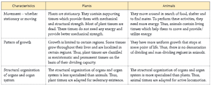
Are plants and animals made of same type of tissues?
If you carefully observe, you will find many differences in plants and animals. The body of the two is specialized to perform specialized functions. Thus, plants and animals have different types of tissues since they differ structurally and perform different functions. Plants are fixed at a place, i.e. they do not move. Therefore, most of the plant tissues are supportive to provide them mechanical strength. Plants also need very little energy and maintenance.
Tissues Class 9 KSEEB notes
Therefore, most of the plant tissues are dead. On the other hand, animals are not stationary at a place. They need a continuous supply of energy and maintenance. Therefore most of the animal tissues are alive and capable of utilizing energy regularly. compares the organization of tissues in plants and animals based on certain characteristics.
Plant Tissues
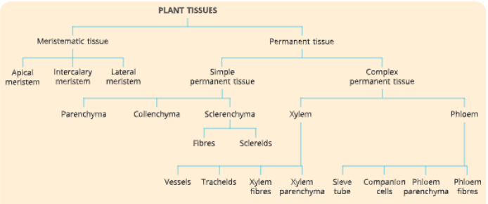
Plant tissues are divided into two types on the basis of their stage of development and dividing capacity These are:
meristematic tissue, and permanent tissue.
A meristematic tissue is a group of young cells that have the capacity of active cell division. Thitissue is found in all the growing regions of a plant, such
as root tip, shoot tip, etc. Let us perform an activity (Activity 1) to learn about meristematic plant tissues.
Characteristics of meristematic tissues They are composed of living cells, and are very active. The cells are thin-walled, small sized and undifferentiated.
Activity 1
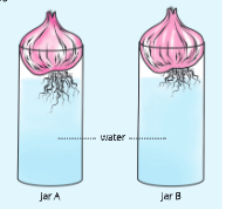
- To show that meristematic tissue is responsible for the growth of onion root tips You will need
Two glass jars, two onion bulbs, water, ruler and razor blade Procedure - Take two glass jars of similar size and label them as A and B. Fill each of these jars with water.
- Take two onion bulbs of similar size and place one on each jar in such a manner that their roots touch the water in the jars.
- Observe the growth of roots in both the onion bulbs for 3 days.
- Measure the length of roots on each day starting from day 1 up to day 3. On day 4, cut the root tips of the onion bulb in jar B by about 1 cm and place the bulbs back in the jar.
- Now, observe the growth of roots in both the jars and with the help of a ruler, measure their lengths each day for five more days. Record your observations in a table as given below:
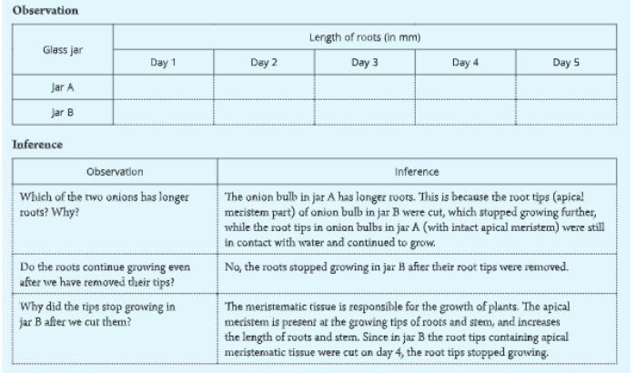
Thus, from the above activity, we can conclude that growth of plants takes place only in certain regions. Meristematic tissues are located in these regions and are responsible for growth.
- The cells have dense granular cytoplasm. The nucleus is large, prominent and centrally located. They have compactly arranged cells without intercellular spaces.
- They are capable of dividing indefinitely, i.e. they have active cell division. New cells produced by meristematic tissues are initially like those of meristem but later as they mature and grow, they become differentiated as components of other tissues.
- They don’t store reserve food material.
- They lack vacuoles.
Types of meristems
According to their positions in the plant body, meristems are divided into three types – apical meristem, lateral meristem and intercalary meristem.
Apical meristem
Apical meristem is found at the growing tips of stem, root and their growing branches. It is also called primary meristem. It consists of a group of cells that give rise to primary permanent tissues that together constitute the primary body of the plant. Due to the growth of apical meristems, there is an increase in the length of stems and roots.
SSLC Biology Chapter 2 Tissues notes
Lateral meristem
Lateral meristem occurs on the sides of roots and stem and is responsible for the increase in the girth (diameter) of the roots and stem. These tissues are also responsible for growth in thickness by the addition of secondary tissue, and this phenomenon is called secondary growth.

It occurs as the cambium of the vascular bundles of dicot roots and sterns and as cork cambium beneath the branch.
Intercalary meristem
This is the part of apical meristem which gets separated from the apex due to the development of permanent tissue in-between. Intercalary meristem helps in the elongation of the organs. It is present mostly at the base of nodes, internodes (space on either side of node) and leaves.
Permenant Tissue
A permanent tissue is a group of cells in which growth has stopped either completely or for the time being. These are formed by cells that have lost the capacity to divide. These cells may be dead or alive, thin- walled or thick-walled. Permanent tissues are formed by the growth of meristematic tissues. The process of taking up a permanent shape, size and function is called differentiation. Permanent tissues are formed by the differentiation of cells of meristematic tissues specialized to perform a particular function. Let us perform an activity to observe various types of permanent tissues in plants.
On the basis of the function performed, permanent tissues can be categorized as
- simple permanent tissues, and
- complex permanent tissues.
Simple permanent tissue (supportive tissue)
The tissues made up of one type of cells, which resemble each other and perform similar function are called simple permanent tissues. These tissues are specialized to perform supportive and protective function. There are three types of simple permanent tissues in plants – parenchyma, collenchyma and sclerenchyma.
Parenchyma
Structure: Parenchyma is a simple, permanent living
Activity 2
To observe various types of permanent plant tissues in the section of a stem You will need A young stem of any dicot or monocot plant, razor blade, glycerine, safranin, slides
and cover slips
Procedure
- Take a young stem of any dicot or monocot plant and with the help of a razor blade cut it into very thin sections. Take the help of your teacher to do this.
- Stain the section of stem with safranin stain. Gently, put one neatly cut section on a glass slide and put a drop of glycerine over it.
- Cover the section with a cover slip and observe under a microscope.
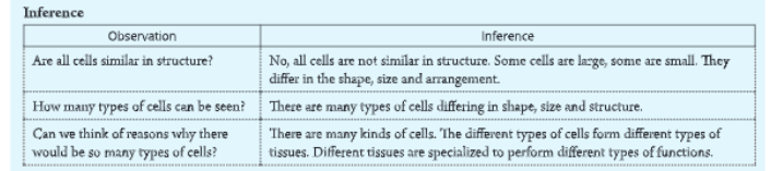
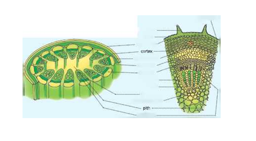 Observation
What do you observe? What kind of cells do you see? Are they similar? How are these cells arranged? Compare your section with the one given here.
 Now, repeat the above activity by taking sections of a plant root or sections of a stem and roots of different plants.
tissue which is made up of unspecialized thin-walled cells . The cells of parenchyma tissue are live and oval, rounded or polygonal in outline.
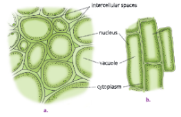 Their wall is made up of cellulose. The cells in these tissues are usually loosely packed. Thus, large intercellular spaces are present in between the parenchyma cells. Each parenchyma cell encloses a large central vacuole, cytoplasm and a prominent nucleus.
Distribution: Parenchyma is found universally in all the plants. It forms the major tissue of softer parts like the epidermis, cortex, pith and leaf mesophyll.
It is also found in xylem and phloem. Functions of parenchyma
- It stores food material in the form of proteins, starch, oil and fats.
- Parenchyma of stems and roots also stores nutrients and water.
- Parenchyma cells provide support and rigidity to the plants by keeping the cells rigid.
- Parenchyma cells form the basic packing tissue and protect the internal tissues.
In the leaves of green plants, parenchyma tissue contains chlorophyll, and is called chlorenchyma.
Chlorenchyma thus helps in photosynthesis.
In many aquatic plants, parenchyma cells have well- developed air spaces and are known as aerenchyma.
These air filled intercellular spaces give buoyancy to plants and help them float in water.
Class 9 Biology Karnataka Board Chapter 2 summary
Collenchyma
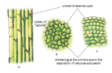
Structure: The cells of collenchyma are living, somewhat elongated with cellulose and pectin thickening at the corners. They are found as longitudinal strips. Collenchyma cells appear circular or oval in transverse section. Internally, each cell possesses a large central vacuole, peripheral cytoplasm and a nucleus.
There is very little intercellular space between cells of collenchyma tissue due to cellulose thickening.
Distribution: Collenchyma tissue is usually found below the epidermis in stem and stalks of leaves (petiole) and midrib of leaves of dicot plants.
Collenchyma is absent in monocot stems.
Functions of collenchyma
- It provides tensile strength and rigidity to the plants due to thickening of the walls.
- Collenchyma also provides elasticity to the plant organs. The flexibility in plants is due to
- collenchyma tissues. It facilitates the bending of leaves and stems without breaking them.
- Collenchyma being alive also stores food.
Sclerenchyma
Structure: It is also a simple permanent tissue.
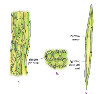
The cells of this tissue are dead. These are long, narrow with tapering ends. Their cell walls are thickened due to lignin which is a chemical substance that acts as a cement and hardens them. Central cavity of the cells is greatly reduced due to this thickening. clerenchyma tissues are of two types-fibres and sclereids.
Distribution: These tissues occur in the veins of
leaves and in hard covering of seeds and nuts. They form the major part of walnut shells and other nuts.
They form an important part of the bark of trees.
Functions of sclerenchyma
- Sclerenchyma provides mechanical strength to the plant and its parts.
- They protect the plant from environmental extremes like strong winds.
- They make the plant hard and stiff. The husk of coconut is made up of sclerenchyma tissue.
Protective plant tissue
In plants, the protective tissues are epidermis and cork.
Tissues Class 9 KSEEB important questions
Epidermis
Let us perform an activity to study the structure of epidermis.
Activity 3
To observe the structure of epidermis from a freshly plucked leaf of Rhoco You will need A freshly plucked leaf of Rhoro, glycerine, safranin, slides, cover slips, microscope
procedure
- Take a freshly plucked leaf of Rhoeo. Clean the leaf gently with water. Stretch the leaf and break it simply by applying pressure. Keep it stretched gently so that some peel projects out from the broken portion.
- Remove this peel and put it in a Petri dish containing water.
- Stain the peel with safranin stain. Let it stain for some time.
With the help of a fine hair brush, gently transfer the stained peel to a glass slide and put a drop of glycerine over it.
Cover the section with a cover slip and observe under a microscope.
Observation
What do you observe? What kind of cells do you see?
Compare your section with the one given in
Inference
The structure you observe is epidermis, the outermost layer of cells.
Characteristics
Epidermis is the outermost protective layer of plant organs. It is usually single-layered but in leaves of some plants growing in dry habitats, it is multi- layered and thick to protect the plant from water loss. The epidermis covers the entire surface of the plant. It protects all parts of the plant. Cells of epidermis are flat and form a continuous layer without intercellular spaces to protect the plant tissues. Outer and side walls of most epidermal cells are thicker than the inner walls.
Distribution
Epidermal cells of aerial parts of the plant secrete a waxy, water resistant layer on their outer surface. It protects them against loss of water, mechanical injury and any attack by pathogens.
In desert plants, outer walls of the epidermis are usually thick and covered with organic substances like cutin. The cutin is a chemical substance that is waterproof. The thick cutinized wall of epidermis greatly reduces the loss of water by transpiration. The epidermal cells of the roots contain long hair-like structures called root hair. The root hairs increase surface area for absorption of water and nutrients from soil.
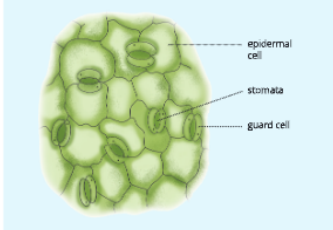
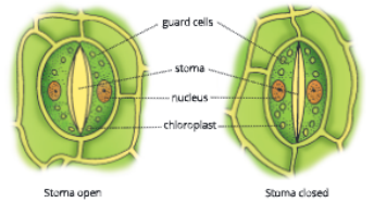
The epidermis of the leaf contains small pores called stomata. Each stoma is enclosed by two kidney. shaped cells called guard cells. The guard cells enclose a central cavity. The stomata help in the exchange of gases with the atmosphere. They also help in the loss of excess water in the form of water vapour, by a process known as transpiration.


Functions of epidermis
- Epidermis performs the function of protection in the following ways:
- It protects internal tissues against mechanical injury; parasitic fungi, bacteria; and cold or heat.
- Thick cuticle, wax, epidermal hair and multiple layers of epidermis reduce the loss of water from the internal tissue.
- Epidermal cells of roots have hair that greatly increase the surface area for the absorption of water and nutrients.
cell body or cyton of another nerve cell. This loose connection between the axon endings of one nerve cell and the cyton of the next nerve cell is called synapse. The other small branches given out by Cork (Phellem) (Gk. phellos: cork) As plants grow older, protective tissues at the periphery undergo certain changes. A strip of secondary meristem replaces the epidermal layer of the stem forming a multiple layered thick bark of the tree called cork. Cork in mature woody stem is made up of dead, thick-walled cells. The cork cells are compactly arranged without any intercellular spaces. The walls of cork cells also contain suberin (a chemical substance) which is impervious to gases and water.
Functions of cork cells Cork performs protective functions in the following ways:
- Cork cells being highly suberized and thick-walled protect the inner tissues.
- Cork provides insulation from freezing temperatures.
- It protects the inner tissues from the attacks of microorganisms and prevents water loss also.
Class 9 SSLC Biology Chapter 2 textbook solutions
Complex permanent tissue (conducting tissue)
Cells of the complex tissues work together as a unit and have a common origin. Complex tissues are made up of more than one type of cells, which work in close coordination to perform a common function. The main complex tissues in vascular plants are xylem and phloem. Both xylem and phloem are assemblage of living and dead cells. They are conducting tissue and together constitute a vascular bundle.
Xylem
Xylem is a complex tissue The cells are thick-walled and many of them are dead. Xylem is mainly concerned with the conduction of water and minerals. It also provides mechanical support to the plant. As a conducting strand, xylem forms a continuous channel through the roots, stem, leaves and other aerial parts. Xylem consists of four types of cells – xylem vessels, tracheids, xylem fibres and xylem parenchyma.
Xylem vessels and tracheids are tubular structures. Tracheids are found in lower vascular plants and gymnosperms but are absent in most angiosperms.
Xylem vessels are found in xylem of angiosperms and are absent in most gymnosperms. Xylem vessels and tracheids help in the conduction (transport) of water and minerals from roots to aerial parts of the plant.

Xylem fibres are found abundantly in woody dicotyledonous plants. They are supporting in nature and provide mechanical strength to the plant body. Xylem parenchyma are the only living components of xylem. They are present in primary and secondary xylem. These are concerned with the storage of food and sideways conduction of water.
Phloem
Phloem is the chief food-conducting tissue of plants. Unlike xylem, materials can move in both directions in phloem. Phloem is responsible for the transport of food prepared by leaves to the other parts of the plant . There are four types of phloem elements – sieve tubes, companion cells, phloem parenchyma and phloem fibres. Except phloem fibres that are dead, all other members of the phloem tissue are living.
Karnataka Board Class 9 Biology Chapter 2 PDF
The sieve tubes of phloem are elongated tubular conducting channels, which are placed end to end. They have perforated walls. They conduct food materials prepared in the leaves and greener young stems to all parts of the plant. Companion cells lie on the sides of sieve tubes and are closely associated with them. They help sieve tubes in the conduction of food materials.
Phloem parenchyma food. parenchyma are ordinary living cells associated with phloem.
They store Phloem fibres are dead sclerenchyma fibres.
They provide mechanical strength. The textile fibres of flax, hemp and jute are phloem fibres.
Animal Tissues
To understand animal tissues let us take an example. When you breathe, your chest moves up and down. The movement in the chest is to accommodate the air (rich in oxygen) that we take in and release the air (rich in carbon dioxide). This movement is brought about by muscle cells in the body. Regular contraction and relaxation of muscle cells bring about this movement.
When you breathe you take in oxygen. This oxygen is transported to the lungs where it is absorbed and is then carried to all parts of the body through blood. Blood also carries food to all the parts of the body. It also collects wastes from various parts of the body and carries them to the liver and kidney for excretion. Thus, in this example, both muscles and blood are examples of tissues. Let us learn in detail about different types of animal tissues.
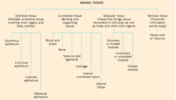
Types of Animal Tissues
On the basis of functions performed by them in our body, there are four major types of tissues in animals.They are
- Epithelial tissue
- Muscular tissue
- Connective tissue
- Nervous tissue
Epithelial Tissue
Epithelial tissue is the simplest tissue. An epithelial tissue is composed of one or more layers of cells covering and protecting the external surface and internal body organs. Location: Epithelium is a protective tissue. It covers most of the organs and cavities of hollow body organs, blood vessels and ducts. It acts as a barrier to keep different body systems separate. The skin, lining of blood vessels, mouth, buccal cavity, pharynx, oesophagus, stomach, lungs and alveoli are lined by the epithelial tissue.
Characteristics of epithelial tissue
- The cells of epithelial tissue are tightly-packed (without any intercellular spaces) and form continuous sheets.
- Cells are cemented together by small amount of viscous cementing substance formed of glycoproteins.
- Cells of the lowermost layer rest on non-cellular gelatinous basement membrane with collagen fibres which separates it from the underlying connective tissue.
Each epithelial tissue is separated from the underlying tissue by a basement membrane.
- The epithelial tissue does not have blood vessels. Functions of epithelial tissue Epithelial tissue performs the following functions:
- Protection: Epithelial tissue protects the underlying tissues from mechanical injury, entry of germs, drying up and harmful chemicals.
- Absorption: Epithelial lining of intestine absorbs water and nutrients from digested food.
- Excretion: Epithelial lining of uriniferous tubules (nephron) in kidneys helps in the excretion of nitrogenous waste.
- Secretion: Epithelial lining of digestive glands and endocrine glands secretes useful secretions.
- Exchange of materials: The cells of various epithelia regulate the exchange of materials between the body and the external environment and also among different parts of the body.
- Epithelial lining of alveoli (lungs) brings about exchange of oxygen and carbon dioxide between blood and inhaled air.
- Barrier: It acts as a selective barrier to anything entering or leaving the organ.
Types of epithelial tissue
Based on the structure and organization of the component cells, the epithelial tissue may be classified as follows:
- Squamous epithelium
- Columnar epithelium
- Cuboidal epithelium
- Glandular epithelium
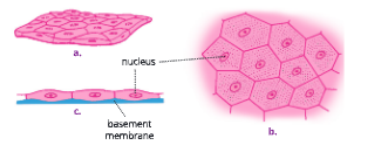
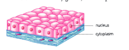
Squamous epithelium
It is formed by flattened, scale-like polygonal cells closely fitted together like tiles in a mosaic floor.
Location:
The squamous epithelium forms the lining of the blood vessels, oesophagus, mouth, nose, skin, alveoli of lungs, etc. . Skin is also made up of squamous epithelium.
Function
It protects the underlying body parts from mechanical injury, germs, drying up, etc.
There are two types of squamous epithelium simple squamous epithelium and stratified squamous epithelium.
Simple squamous epithelium:
It is made up of extremely thin and single layer of simple flat cells that form a delicate lining. It lines the blood vessels, nose, coelomic cavity, bronchioles or alveoli in lungs, where transportation of substances occurs through selectively permeable membrane.
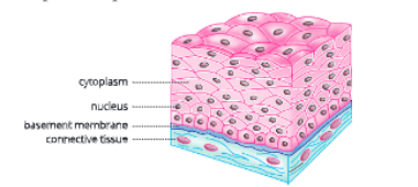
Stratified squamous epithelium:
It contains cells arranged in a pattern of layers to prevent any wear and tear. For example, skin epithelial cells are arranged in a pattern of many layers of stratified squamous epithelium.
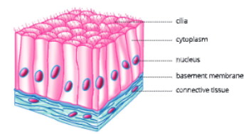
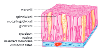
Columnar epithelium
Columnar means pillar-like. Therefore, as the name suggests, it is formed of tall pillar-like cylindrical cells

lying side by side. They appear polygonal in shape in the surface view. The cells are much taller than they are wide. The basal part of cells bears oval nuclei .
Location:
The columnar epithelium is found in organs where absorption and secretion occur like inner lining of intestine, pharynx, larynx and oviduct. It facilitates the movement across the epithelial barrier. It is present in the walls of stomach, intestines and the gall bladder. It is also present in gastric and intestinal glands.
Functions:
The columnar epithelium helps in absorption through the lining of stomach and intestine and helps in the secretion of mucus through the goblet cells or mucous membrane. In the respiratory tract, there are simple hair-like projections called cilia or microvilli or brush border on the outer surfaces of columnar epithelial cells. These cilia or brush border can move and push the mucus forward to clear it of any unwanted particles like dust. This is known as ciliated columnar epithelium.
Cuboidal epithelium
It is made up of cube-shaped cells of almost equal height and width. In the surface view, they look polygonal in shape. The nuclei are round in shape and lie in the centre of the cells.
Location:
It is present in the lining of kidney tubules and ducts of salivary glands, where it provides mechanical strength.
It also lines sweat glands, thyroid glands and germinal epithelium of testes and ovaries.
Function:
It helps in absorption, excretion as well as secretion other than providing mechanical support.
The Fundamental Unit of Life and Tissues Class 9 notes KSEEB
Glandular epithelium
Often, epithelium specializes to form glands. Glands develop from epithelial tissue which can secrete substances at the epithelial surface. Sometimes there is inward folding of epithelial tissue forming multicellular gland, called glandular epithelium.
Location:
Goblet cells in the mucous membrane of alimentary canal, sweat glands and sebaceous glands in the skin, mammary glands, salivary glands in the mouth, etc.
Functions:
Glandular epithelium is a modified columnar epithelium. Cells of glandular epithelium are modified and specialized to secrete certain substances. Glandular epithelium helps in secretion of hormones, sweat, saliva, digestive enzymes, etc.
Connective tissue is a binding and supporting tissue.
Characteristics of connective tissue
Cells of connective tissue are loosely spaced and embedded in an intercellular matrix. Basically, connective tissue consists of matrix, connective tissue cells and connective tissue fibres. Matrix is homogeneously fibrous in nature and binds other tissues. It is also called packing tissue. It is non-living and an amorphous, transparent substance. It helps in the diffusion of food materials, water and gases across the cells.
Location:
It is distributed throughout the body and forms about 30% of the body weight. It forms a sheath around the organs.
Functions of connective tissue
- Binding and packing:The main functions of connective tissue are binding, supporting and packing different organs of the body together.
- Attachment: Connective tissue binds different organs with one another, for example, muscles with skin, muscles with bones.
- Support: It forms a supporting framework of cartilage and bones in the body.
- Storage: Adipose connective tissue helps in the storage of fats. It also forms shockproof cushions around kidneys, ovaries and eyeballs
- Protection: It forms protective sheaths around delicate organs such as spleen, kidneys, testes, etc.
- Defence: White blood corpuscles and lymph act as phagocytes and provide protection against bacterial infections.
- Repair: Collagen fibres of connective tissue help in the repair of injured tissues.
Types of connective tissue There are following types of connective tissues in the human body
- Fluid connective tissue – blood and lymph
- Skeletal tissue – bone and cartilage
- Fibrous connective tissue-tendons and ligaments
- Areolar connective tissue
- Adipose tissue
Fibrous connective tissue, areolar tissue and adipose tissue are together called connective tissue proper.
Fluid connective tissue -blood and lymph
Both blood and lymph are fluid connective tissues. and hormones to various cells and tissues and also remove CO₂ and other waste from the cells. They have a fluid matrix. Blood cells cannot divide. Blood consists of blood corpuscles suspended in 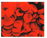 blood plasma. Plasma is a straw-coloured fluid which contains water, inorganic salts, organic substances like blood proteins and hormones. Red blood cells (RBCs), white blood cells (WBCs)
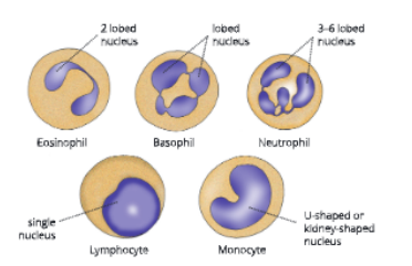
and platelets are suspended in the plasma. RBCs and WBCs are living while platelets and plasma are non-living.
Lymph is a transparent, light yellow fluid. It is not red in colour due to the absence of RBCs. It contains white blood corpuscles called leucocytes. Lymph is present in the intercellular spaces, hence it is also called tissue fluid.
WBCs act as phagocytes and remove any foreign elements in the blood, thereby providing protection against bacterial infection.
Functions:
Blood flows to all parts of the body and connects different parts of the body. Blood plasma transports gases (oxygen and carbon dioxide), digested food, hormones and waste materials to different parts of the body. Prothrombin and fibrinogen present in plasma help in the clotting of blood.
Skeletal tissue – bone and cartilage
The bone and cartilage form skeletal tissue. Skeletal tissue forms the endoskeleton of vertebrate body. Bone Bone is a rigid (non-flexible) and hard skeletal connective tissue. It forms the skeletal framework that supports the body. It also supports the muscles and main organs of the body. Its matrix is hard. The bone cells are called osteocytes. Bone cells are embedded
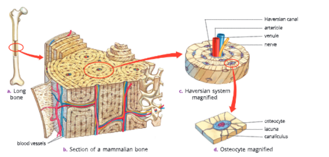
in a hard matrix which is composed of calcium and phosphorus compounds. Each bone cell is enclosed in a small fluid-filled ring-shaped cavity called the lacuna. The osteocytes are present in the concentric rings, called lamellae, around the central canal. This central canal is called the Haversian canal. Canaliculus contains slender process of bone cells or osteocytes.
Cartilage
Cartilage is a compact, relatively soft and elastic skeletal tissue. It consists of elastic matrix having proteins (condrin), collagen fibres, sugar and is slightly hardened by calcium. Cartilage cells, called chondrocytes, are present in fluid-filled spaces called lacunae. Cartilage has widely spaced out cells. In human beings, cartilage is present in the larynx, trachea, at the end of bones, nose and in between ribs and sternum.
Functions:
Cartilage smoothens bone surface at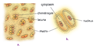
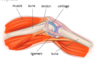
joints and provides support and flexibility to the body parts where it is found. It prevents wear and tear of long bones, where it is present at the end. Fibrous connective tissue – tendons and ligaments (L. tendo: to stretch) Tendons are dense fibrous connective tissues with great strength and flexibility. These occur in the form of tendons or sheets. Their matrix contains unbranched white collagen fibres. Tendons join muscles with bones.
Ligaments contain very little matrix with many closely-packed yellow or elastic fibres. Due to the presence of yellow elastic fibres in matrix, they are very elastic and connect one bone with the other. They have extensive strength and facilitate bending and rotational movement of bones over joints.
Areolar connective tissue
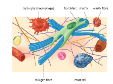
It is the simplest and most widely distributed connective tissue in the body. In this tissue, fibres are loosely arranged in a meshwork. Its matrix is jelly-like and contains large star-shaped
fibroblast cells or fibrocytes, irregular mast cells, white collagen fibres, yellow elastic fibres, lymphoid cells and large histiocytes . Fibrocytes secrete fibres.
Function:
Areolar connective tissue binds the skin with muscles and attaches blood vessels and nerves to the surrounding tissues. It fills the space inside the organs and supports internal organs. It also helps in the repair of tissues. Overall, it acts as a supporting and packing tissue among organs lying in the body cavity.
SSLC Class 9 Biology Tissues chapter worksheet
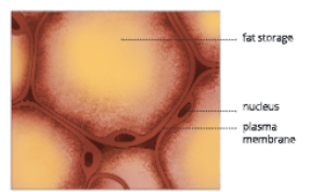
Adipose tissue
It is an aggregation of fat cells called adipocytes. It is found below the skin and between internal organs, around kidneys and in yellow bone marrow. The cells of adipose tissues are filled with fat globules. Thus, it stores fat which acts as an insulator. It also acts as a cushion for shock absorption.
Muscular Tissue (the Contractile Tissue)
The muscle tissue consists of long, narrow cells called muscle fibres. The adjacent muscle fibres are held together by connective tissue. Muscles bring about movement of body parts and locomotion in organisms.
The cytoplasm of muscle fibre is called sarcoplasm. It is a highly contractile myofibril. Myofibrils contain contractile proteins actin and myosin which contract and relax to cause movement.
Types of muscular tissue
In human beings, three types of muscles are present – voluntary muscles, involuntary muscles and cardiac muscles.
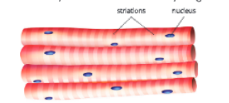
Voluntary or striated muscles
A voluntary or striated muscle consists of cells with long, narrow, cylindrical and unbranched fibres with blunt ends. These muscles when stained show alternate dark and light bands or striations . Each muscle fibre is multinucleated (has many nuclei).
Location:
Striated muscles are present in the body wall, limbs, tongue, pharynx and at the tip of oesophagus.
Functions:
These muscles can be moved at our will. As their movement is under our will, they are popularly called voluntary muscles. These muscles are also called skeletal muscles as these are attached to bones. They form almost 50% of body weight.
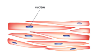
Involuntary or Unstriated Muscles
These muscles are also called smooth muscles as they lack transverse striations. These muscle cells are spindle- shaped (long with pointed ends) and are arranged in bundles. They have only one nucleus (uninucleated). They are also called unstriated muscles as they do not contain any striations or bands.
Location:
These muscles are found in the iris of eye, in ureter and in the bronchi of lungs.
Functions:
Their movement is not under our will and hence these are called involuntary muscles. We cannot start or stop their action.
Cardiac muscles
Structurally, these muscles are cylindrical, branched and uninucleated . They form an interconnecting network. The muscle filaments are connected by dark junctions called intercalated disc. These act as impulse boosters.
Karnataka State Board Class 9 Biology Chapter 2 explanation
Location
These muscles are exclusively present in the heart.
Functions:
These muscles work rapidly, rhythmically and tirelessly, contracting and relaxing endlessly from early embryonic stage until death.
Nervous Tissue
Nervous tissue consists of nerve cells or neurons. They are specialized to respond to stimuli and transmit stimulus very rapidly from one part to another within the body. The brain, spinal cord and nerves are composed of nervous tissue. A neuron or nerve cell is the structural and functional unit of the nervous system. A typical nerve cell consists of cell body or cyton, axon, dendrons and dendrites. Cell body or cyton is star-shaped and has a prominent nucleus and cytoplasm. Its cytoplasm contains a large nucleus, fine thread-like neurofibrils and Nissl granules. From the cell body arise several branches. One of the branches grows very large in comparison to others. This branch is called axon. The axon terminates into axon endings.
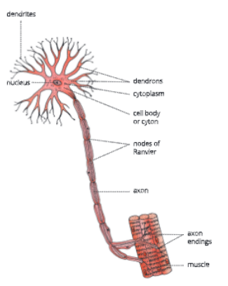
The axon endings of one nerve cell is loosely-placed on the cyton are called dendrons, which further divide to form dendrites. The dendrites receive impulses and axon takes the impulses away from the cell body or cyton. Both nerve and muscle tissue work in coordination and help animals to move in response to stimuli.
Summary
- Tissue is a group of cells similar in structure and function.
- The microscopic study of tissues and their functions is known as histology.
- Plant tissues are of two types – meristematic tissue and permanent tissue.
- Meristematic tissue is dividing tissue present in the growing regions of plants such as root apex and stem apex.
- According to their positions in plants, meristems are divided into apical meristem, intercalary meristem, and lateral meristem.
- A permanent tissue is a group of cells in which growth has been either stopped completely or for the time being.
- Permanent tissues are derived from meristematic tissue once they have lost the ability to divide. Permanent tissues are classified as simple and complex tissues.
- Parenchyma, collenchyma and sclerenchyma are three types of simple permanent tissues.
- Epidermis and cork are the protective tissues in plants.
- A group of more than one type of cells working together as a unit and having a common origin is called a complex tissue. Xylem and phloem are complex permanent tissues.
- Xylem is a complex plant tissue. Its components are xylem vessels, tracheids, xylem fibres and xylem parenchyma.
- Only xylem parenchyma Is the living component.
- Phloem is a chief food-conducting tissue of a plant. Its components are sieve tubes, companion cells, phloem parenchyma and phloem fibres. Except phloem fibres, all other components are living.
- There are four major types of animal tissues – epithelial, connective, muscular and nervous.
- In epithelial tissue, cells are closely-packed and form a continuous sheet. The cells of epithelial tissue rest on basement membrane. Depending upon shape and function, epithelial tissue is classified as squamous, cuboidal, columnar and glandular.
- Connective tissue is a binding and supporting tissue. It forms about 30% of the body weight. In our body, connective tissue includes blood, lymph, bone, cartilage, tendons, ligaments, areolar tissue and adipose tissue.
- Bone cells are star-shaped and are called osteocytes. In mammalian bone, the bone cells are present in concentric rings around the Haversian canal.
- Cartilage cells are present in fluid-filled spaces called lacunae.
- Striated, unstriated and cardiac muscles are the three types of muscle tissues.
- The muscular tissue consists of long narrow cells called muscle fibres which are held together by connective tissue.
- Smooth muscles and cardiac muscles are involuntary. Their movement is not under our will whereas striated muscles are voluntary muscles. Their movement is under our will.
- Structurally, cardiac muscles resemble skeletal muscles and functionally, these are similar to smooth muscles.
- Nervous tissue consists of nerve cells. Each nerve cell consists of cell body or cyton, axon, dendrons and dendrites.
Tissues Class 9 KSEEB question answers
Key Terms
- Histology: The microscopic study of tissues and their functions
- Stomata: The small pores present on the leaf epidermis that help in the exchange of gases with the atmosphere
- Tendon: The fibrous tissue that connects muscles with bones
- Ligament: The fibrous tissue that connects one bone with the other
- Lacuna: A small cavity that encloses bone cells
- Osteocytes: The bone cells
- Chondrocytes: The cartilage cells
- Voluntary muscles: The muscles that can be moved at our will
- Involuntary muscles: The muscles that cannot be moved at our will
- Neuron: The structural and functional unit of the nervous system