
KSEEB Solutions for Class 8 Biology Chapter 6 Circulatory System Notes
All living organisms need food and minerals for survival. In both the animals and the plants, there is a transport system that carries the food and other substances to the various parts of the body. This transportation system in the animals and human beings is called the circulatory system.
The main transport system in human beings is the ‘blood circulatory system’ (which is commonly known as just ‘circulatory system’). In the circulatory system, blood carries digested food, water and oxygen to all the parts of the body.
It also takes away the waste products like carbon dioxide made in the body cells. Thus, the blood circulatory system makes food, water and oxygen available to every part of the body, and helps in removing waste materials of the body like carbon dioxide, etc.
Kseeb Class 8 Biology Chapter 6 Circulatory System Solutions
Circulatory System
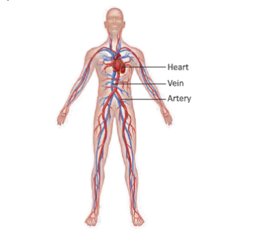
The various organs of the circulatory system in humans are: Heart and Blood vessels (Blood vessels are of three types: arteries, veins and capillaries). Blood is also considered a part of the circulatory system. In the circulator)’ system, the heart acts as a pump to push out blood. The blood vessels (arteries, veins and capillaries) act as tubes or pipes through which blood flows in the whole body.
Sslc Class 8 Biology Circulatory System Question And Answers
Blood
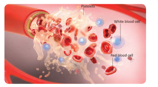
Blood is a red coloured liquid which flows in blood vessels and circulates in our body. Blood is red because it contains a red pigment called haemoglobin. Blood has many different cells which perform different functions. Blood consists of four components: plasma, red blood cells, white blood cells and platelets.
Plasma is a liquid and red blood cells, white blood cells and platelets keep floating in it. The blood is able to transport (or carry) various substances in the body due to the presence of different components in it. We will now describe all the four components of blood and their functions in somewhat detail.
Plasma
The liquid part of blood is called plasma. Plasma is a pale yellow, sticky liquid. It is 90 per cent water and 3.5 per cent common salt. Plasma contains dissolved substances such as digested food and waste products (like carbon dioxide and urea). Plasma carries water and dissolved substances such as digested food and waste products from one part to another part in the body.
Kseeb Class 8 Biology Circulatory System Textbook Solutions
Red Blood Cells (RBCs)
Red blood cells are red in colour due to the presence of a red pigment called haemoglobin inside them. Haemoglobin present in the red blood cells carries oxygen to different parts and ultimately to all the cells. If there is a deficiency of haemoglobin in the blood of a person, it becomes difficult to provide oxygen efficiently to all the cells of his body.
Circulatory System Class 8 Biology Kseeb Important Questions
White Blood Cells (WBCs)
The white blood cells fight infection and protect us from diseases. This is because white blood cells help to fight against germs which may enter our bodies and cause diseases. Some white blood cells can eat up the germs (like bacteria) wrhich cause diseases. Other white blood cells make chemicals known as antibodies’ to fight against infection. White blood cells are much smaller in number than red blood cells.
Platelets
Platelets arc the tiny fragments of special cells formed in the bone marrow. Platelets help in the clotting of blood in a cut or wound. When someone gels injured, then blood starts flowing from the cut made by the injury. After some time, however, a dark red clot is formed which plugs the cut and bleeding stops.
If, however, there were no platelets in the blood, then bleeding caused by a cut from an injury would not stop. This may cause loss of too much blood from the body of a person leading to death.
Karnataka Sslc Class 8 Biology Chapter 6 Solutions In English
Functions of Blood
The blood performs number of important functions in our body. Some of these functions have been discussed here:
- Blood helps in carrying oxygen and carbon dioxide between the respiratory organs and tissues.
- Blood helps in carrying the stored food and vitamins to the tissues or organs where they are generally needed.
- It helps in transporting the waste products from the tissues to the excretory organs and regulates the water balance.
- It contains antibodies to fight against the infections in the body.
- It helps in transporting the nutrients and water to all parts of the body from the alimentary canal.
- It prevents excessive bleeding in case of accidents by forming blood clots.
- It carries hormones from the glands to the parts where it is needed for action.
- It helps in regulating the temperature of an organism and distribution of heat equally in all parts of the body.
Blood Groups
Blood type am vary from person to person. The red blood cells in the blood contain number of protein molecules called the antigens. The antigens are foreign substances in the body of humans that initiate the production of antibodies by the human immune system. The antibodies are chemical substances which are made by the human body in response to the foreign substances or antigens.
Thus, based on the antigens and antibodies, blood group also differ. Karl Landsteiner discovered that sometimes during blood transfusion from one person (donor) to the other (recipient), the foreign blood lends to clump and cause shock or jaundice.
This happens when the blood groups of the two human beings, the donor and the recipient, are different. Hence it is very important to know the blood groups of different people before any such process. The blood groups are differentiated on the basis of antigens and antibodies present in the blood.
- The antigens are found on the surface of RBCs and the antibodies are present in the plasma.
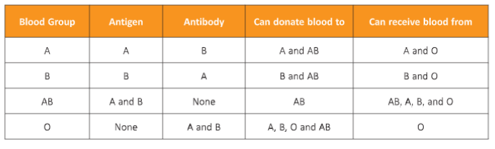
- There are four types of blood groups (A, B, AB, and O) in human beings based on the presence or absence of antigens and antibodies.
- The blood group A has antigen A on its RBCs and antibody B.
- The blood group B has antigen B and antibody A.
- The blood group AB has both antigens A and B but no antibodies.
- The blood group O has no antigens but both antibodies A and B.
- O type blood can be given to persons of all type of blood groups such as O, A, B, AB. The person having blood group O is called Universal donor. The person with blood group AB can receive the blood from all type of blood groups, Le. AB, A, B, and O and is therefore called Universal acceptor.
Kseeb Class 8 Biology Circulatory System Solved Exercises
Blood Transfusion
The transfer of blood from one person to the other is called blood transfusion. When people are seriously ill or get injured then, there are chances that they lose a lot of blood and thus, might need blood transfusion. The person who receives blood is called recipient. The person who donates blood is called donor. It is necessary to match the type of blood of the recipient and the donor before transfusion.
The antibodies and antigens present in blood act against each other. When the blood of opposite antigen is transfused into the body of a person, the antibodies of the person act against the antigens and cause clumping of the blood. Tt may lead to infections or even death. Thus, blood transfusion must be done by matching the blood groups of donor and recipients.
Class 8 Biology Circulatory System Notes Karnataka Board
Rhesus or the Rh factor
The blood contains another important antigen which is Rh factor or the Rhesus factor. This determines the compatibility of the blood transfusion to be given to the recipient. The Rh factor can be positive or negative. A person can be called Rh positive or Rh negative, depending on the presence or absence of the Rh factor.
The Rh negative people do not have an antibody in the plasma as against the Rh factor. If a person with Rh negative is given blood with Rh positive by mistake, then recipient develops antibodies against the Rh factor. Repeated transfusion of blood to the recipient with Rh positive factor can become very dangerous and may endanger the recipient’s life.

Blood Vessels
Blood vessels carry blood throughout our body. ’Ihe blood vessels run between the heart and the rest of the body. There are three types of blood vessels in our body—arteries, veins and capillaries.
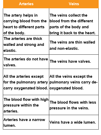
Arteries
Arteries are the blood vessels which carry blood from the heart to all the parts of the body. In other words, arteries are the blood vessels which carry blood away from the heart. Arteries arc found in the whole of our body. The arteries have thick and elastic walls, because blood flows through them at high pressure. The main artery (called aorta) is connected to the left ventricle of the heart.
The main artery carries oxygenated blood from the left ventricle to all the parts of the body (except the lungs). Another artery called pulmonary artery is connected to the right ventricle of the heart. The pulmonary artery carries deoxygenated blood from the right ventricle to the lungs.
Circulatory System Class 8 Biology Summary And Explanation Kseeb
Veins
Veins are the blood vessels which carry blood from all the parts of the body back to the heart. In other words, veins are the blood vessels which carry blood towards the heart. The greenish-blue lines which we see just below the skin on our hands and legs are the veins.
The deoxygenated blood returns to the heart at low pressure through the veins. Since the blood flows at low pressure through the veins, so the veins have thin walls.
Veins have valves in them which allow the blood in them to flow in one direction only towards the heart. The valves prevent the backflow of blood in veins. The main vein is connected to the right atrium of the heart. The main vein carries deoxygenated blood from all the parts of the body (except lungs) back to the heart.
Another vein called pulmonary vein is connected to the left atrium of the heart. The pulmonary vein carries oxygenated blood (rich in oxygen) from lungs back to the heart.
Sslc Class 8 Biology Chapter 6 Workbook Answers
Capillaries
Capillaries are the extremely thin blood vessels which connect arteries to veins. Thus, capillaries are always present in-between the arteries and veins in our body. Capillaries (also called ‘blood capillaries’) are present throughout our body. Every cell of the body is near a capillary. The capillaries have extremely thin walls which allow substances to pass from blood into the body cells, and also from body cells into the blood.
The oxygenated blood from arteries enters into the capillaries in all the parts of the body. The various dissolved substances present in the blood (like food and oxygen) pass into body cells through the thin walls of the capillaries. At the same time, the waste products (like carbon dioxide) formed in the body cells enter into blood through the thin walls of capillaries.

Heart
The heart is an organ which pumps blood to all the parts of our body through a network of tubes called blood vessels. Our heart ‘beats’ continuously to circulate blood in the body. The heart works like a pump non-stop throughout our life. The heart lies between the two lungs and above the diaphragm in the chest cavity. The heart is made of special muscle called ‘cardiac muscle’.
The heart is surrounded by a two-layered tissue membrane called pericardium. The space between the two layers is filled with fluid called pericardial fluid.
Structure of Heart
Human heart has four compartments called ‘chambers’. The upper two chambers of heart are called atria (singular of atria is atrium), and the lower two chambers of heart are called ventricles. On the left side of the heart are left atrium and left ventricle. On the right side of the heart are right atrium and right ventricle.
These chambers are meant to prevent the mixing of pure and impure blood. Right and left ventricles are separated by an interventricidar septum.
Kseeb Class 8 Biology Circulatory System Mcqs With Answers
Auricles
Auricles (or atrium) are the upper chambers of the heart. They have thin walls and receive blood from different parts of the body. The right auricle receives the impure blood while the left auricle receives the pure blood.
Ventricles
The lower chambers of the heart are known as ventricles. They have thick walls as they have to pump the blood out of the heart to different parts of the body. The right ventricle pumps the impure blood while the left ventricle pumps the pure blood A thick muscular septum wall is present between two auricles and the two ventricles.
The septum divides the heart into left and right sides.

Class 8 Biology Kseeb Circulatory System Short And Long Answer Questions
Heart valves
Valves inside heart regulate the flow of the blood. They don’t allow oxygenated blood to mix with deoxygenated blood. The right auricle opens into the right ventricle through the auriculo-ventricular opening which is protected by a tricuspid valve. It helps prevent back flow of blood into right auricle. Similarly, a bicuspid valve at the opening of the left auricle into ventricle prevents back flow of blood.
At the base of pulmonary artery and the aorta are the semi-lunar valves which direct the flow of blood and prevents back flow.
The valves present in the heart are discussed here:
- Tricuspid valve: It is located at the opening between right auricle and right ventricle.
- Bicuspid or mitral valve: It is located between left auricle and left ventricle.
- Pulmonary semilunar valve: It is present at the opening of right ventricle into pulmonary artery.
- Aortic semilunar valve: It is located at the point of origin of aorta from left ventricle.
- Superior vena cava: It brings deoxygenated blood from upper body parts (head, neck, chest and arms) to the right auricle.
- Posterior vena cava: It brings deoxygenated blood from posterior or lower body parts, i.e. abdomen and legs to the right auricle. It is the largest vein.
- Aorta: It arises from left ventricle and carries oxygenated blood to supply it to all body parts. Abdominal aorta is the largest artery.
- Coronary arteries: There are two coronary arteries right and left, arising from the base of aorta and supply blood to heart muscles.
Blood Circulation
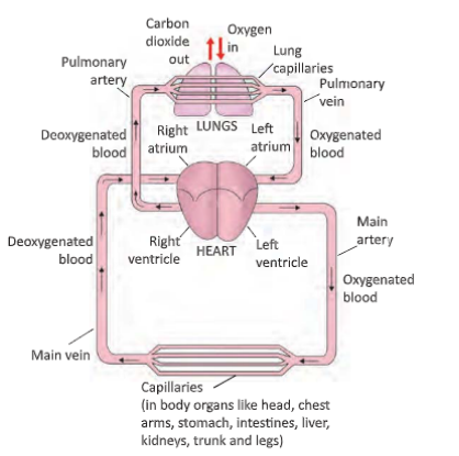
The blood circulates in our body by the pumping action performed by the heart. The pumping action of heart starts by the contraction of its muscular walls. The alternate contraction and relaxation continues regularly. The blood circulates twice through the heart making one complete round of blood circulation through the body.
This is called double circulation. In this arrangement, blood flows between lungs and heart and then between heart and body. That way in the human beings, the circulation cycle works in two ways: pulmonary (lung) circulation and systemic (body) circulation.
Pulmonary Circulation
In this process, the right ventricle sends de-oxygenated blood to tire lungs for oxygenation through pulmonary artery and the left auricle receives oxygenated blood from the lungs through pulmonary vein.
Systemic Circulation
In this process, the right auricle receives de-oxygenated blood from the different parts of the body and the left ventricle sends oxygenated blood through aorta to different parts of the body. In blood circulation, the de-oxygenated blood is collected from the different parts of our body in the pulmonary artery through two major veins called the vena cava and is brought to the right auricle. After the contraction of the right auricle, the blood is pushed into the right ventricle.
When the right ventricle contracts, then the tricuspid valve prevents the blood from flowing back into the right auricle. From the right ventricle, the blood gets pumped into the pulmonary artery which carries the blood into the lungs. There is an exchange of gases, i.e., oxygen and carbon dioxide in the lungs. The oxygenated blood is then carried into the left auricle by the help of the pulmonary vein. Left auricle pumps the blood into the left ventricle. The left ventricle opens into the large artery called the aorta which carries blood out to different parts of the body.
Class 8 Kseeb Biology Structure Of Heart And Blood Vessels Notes
Heartbeats
The heart pumps blood into arteries by contracting. When the heart contracts, it becomes smaller in size and pushes the blood into main artery with a great force. Then the heart relaxes (comes back to its original size) and gels filled up with blood from pulmonary vein. In this way, the heart keeps on contracting and relaxing again and again to pump blood into the body continuously. One complete contraction and relaxation of the heart is called a heartbeat.
The heart of an adult person usually beats about 72 to 80 times in a minute (while resting). This is called the heart rate. We can feel our heartbeats if we place our hand on the left side of our chesl just above the heart region. The heart beats can be counted easily by counting the pulse.
Though the average number of heartbeats of an adult person while resting is about 72 to 80 beats per minute but the number of heartbeats increase during and after a physical exercise or when a person is excited. The heart beats faster during and after a physical activity or exercise because our body needs more energy under these conditions.
The faster beating of heart pumps blood more rapidly to the body organs which supplies more oxygen to the body cells for rapid respiration to produce more energy. The increase in number of heartbeats caused by exercise, excitement, fear or nervousness, however, lasts for a short time after which it becomes normal. The average heart rate in infants is far higher than in adults. A doctor listens to our heartbeats with the help of an instrument called stethoscope.The stethoscope amplifies (makes louder) the sound of heartbeats so that the doctor can hear the heartbeats clearly.
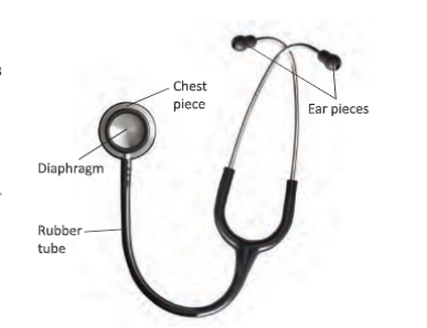
A stethoscope consists of three parts:
- A chest piece (which carries a sensitive diaphragm at its bottom). The diaphragm amplifies the soimds of heartbeats.
- Two ear pieces (which are made of two metal tubes). These are put by the doctor into his ears.
- A rubber tube which joins the chest piece to the ear pieces. The rubber tube transmits the sound from the chest piece into the ear pieces. A stethoscope is used to hear clearly the ‘heartbeats’ by placing the chest piece over the heart region of chest. The diaphragm amplifies the sounds of heartbeats coming from within the body and the rubber tube and ear pieces transmit these sounds to the ears of the doctor. Doctors can get clues about the condition
of our heart by listening to the heartbeats through the stethoscope.
Pulse
Every time the heart beats, blood is forced into arteries. This blood makes the arteries expand a little. The expansion of an artery each time the blood is forced into it, is called pulse. Each heartbeat generates one pulse in the arteries, so the pulse rate of a person is equal to the number of heartbeats per minute i.e., 72 to 80 per minute. Thus, the pulse rate is the same as the heart rate. Just like heartbeats, the pulse rale of a person is higher after a physical exercise or when a person is excited.
Most of our arteries lie deep inside our body and hence cannot be used to feel the pulse. But the wrist, temple and neck are some places where the arteries are close to the surface of skin and we can feel the pulse with our finger tips. The pulse is traditionally taken above the wrist. We usually see the doctor taking the pulse rate of a patient by keeping his fingers on the wrist of the patient and at the same lime looking into his watch.
Blood Pressure
The pressure at which blood is pumped around the body by the heart is called blood pressure. The blood pressure of a person is always expressed in the form of two values called ‘systolic pressure’ and ‘diastolic pressure’. The phase of the heart beat when the heart contracts and pumps the blood into arteries is called ‘systole’. And the phase of heart beat when the heart relaxes (or expands) and allows the chambers to till with blood is called ‘diastole’.
The maximum pressure at which the blood leaves the heart through the main artery (aorta) during contraction phase, is called the systolic pressure. This high pressure in the main artery maintains a steady flow of blood in all the arteries towards the capillaries.
The minimum pressure in the arteries during the relaxation phase of heart is called the diastolic pressure. The value of diastolic pressure is always lower than that of the systolic pressure. The blood pressure of a person is expressed in terms of millimetres of mercury (which is written as mm Hg). The normal blood pressure values arc:
Systolic pressure: 120 mm Hg
Diastolic pressure: 80 mm Hg
This is usually written as 120/80.
Karnataka Board Class 8 Biology Circulatory System Diagram And Explanation
Conditions Related To The Heart Functions
Heart is a very sensitive organ. Here are some of the conditions that may result from the improper functioning of the heart. Some of them have been discussed here.
Palpitation
You must have felt racing heartbeats in a nervous situation such as during exams. This condition arises when the heart beats very fast and we feel the beating of our own heart. The person feels breathlessness, tightness around the chest and dizziness also. This condition is called palpitations.
Cardiac Arrest
Another condition related to the functioning of heart is cardiac arrest. The person collapses and loses consciousness. Cardiac arrest or heart attack occurs when there is an obstruction in the flow of the blood, in arteries. This may happen due to a clot or thickening of the arteries due to cholesterol deposition. It could also happen due to damage in coronary arteries. High blood pressure, obesity, diabetes, smoking or drinking too much alcohol are some reasons behind this condition of heart.
Hypertension
When a person is having higher than normal systolic and diastolic pressure, the person is said to be suffering from hypertension or high blood pressure. This condition prevails even if a person is at rest. Hypertension is the major cause of diseases such as diabetes and heart diseases.
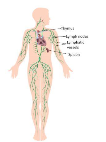
Lymphatic System
A system of tiny tubes called lymph vessels and lymph nodes (or lymph glands) in the human body which transports the liquid called lymph from the body tissues to the blood circulatory system is called lymphatic system. Lymph capillaries are tiny tubes which are present in the whole body just like blood capillaries. Since the ends of the lymph capillaries in the body tissues are closed, so the tissue fluid can only seep into the walls of the lymph capillaries present in the body tissues.
Moreover, since the pores in the walls of the lymph capillaries are somewhat bigger, so even large protein molecules present in the tissue fluid can enter into lymph capillaries. The lymph capillaries join to form larger lymph vessels. The lymph vessels have lymph nodes (or lymph glands) at intervals. The lymph nodes contain special type of cells called lymphocytes. Lymph nodes containing lymphocytes are involved in the cleaning of lymph and protecting the body from disease.
The lymph vessels are connected to large veins of the blood circulatory system. Thymus and spleen are important lymphatic organs. Lymph is a light yellow liquid which is somewhat similar in composition to blood plasma. It contains large protein molecules and digested food (which come into it from the tissue fluid between the cells). It also contains germs from the cells and fragments of dead cells.
Lymph is another medium of circulation in the human body. But lymph flows in only one direction – from body tissues to the heart. Since lymph is derived from the tissue fluid which remains outside the cells of the body, so it is also called extracellular fluid. Lymph contains a special type of white blood cells called lymphocytes which help in fighting infection and disease.
Lymph containing large protein molecules, digested fat, germs and fragments of dead cells from the tissue fluid around the body cells seeps into the lymph capillaries present throughout the body. From lymph capillaries, lymph passes into larger lymph vessels containing lymph nodes.
In the lymph nodes, lymph is cleaned by white blood cells called lymphocytes. These white blood cells eat the germs and dead cells, and also make antibodies for protecting the body from disease.
The cleaned lymph containing large protein molecules, digested fat and other useful materials is transported by lymph vessels to the large veins (called subclavian veins) which run just beneath the collar bone. These veins carry the lymph to the heart. In this way, the circulation of lymph from the body tissues to the heart is completed.
The Functions of Lymph (or Lymphatic System)
- Lymph (or lymphatic system) takes part in the nutritive process of the body. It puts into circulation large protein molecules and digested fat by carrying them from the tissues into the blood stream.
- It protects the body by killing the germs drained out of the body tissues with the help of lymphocytes contained in the lymph nodes, and by producing antibodies.
- Lymph (or lymphatic system) helps in removing the waste products like fragments of dead cells, etc.
Activity
Aim: To measure the pulse and heart rate Material Required: Stopwatch Procedure:
- Work in pairs.
- One student of each pair will measure the pulse rate and heart rate of another student in the pair.
- The first student will place his middle and index fingers on the inner side of the other student s wrist. There will be a regular thumping in the wrist. This thumping is pulse.
- Count the pulse for 30 seconds and multiply it by 2. This gives average pulse rate in one minute.
- Now count the pulse rate after a jog. Repeat the process with other student of the pair.
- Observations: The pulse rate at rest is and after physical exercise is .
- Inference: The pulse is less at resting stage comparec to that after physical exercise. This is because exercise increases the body’s need of oxygen which in turn increases the pulse rate.
Keywords
- Circulatory system: Basic transport system of food ard other substances in the animals and human beings
- Artery: Vessel which carries oxygenated blood to various body tissues
- Veins: Vessels which carries deoxygenated blood to heart
- Haemoglobin: Red colour pigment present in the blood
- Diastole: A term used when the heart is relaxed
- Systole: A term used for contraction of the heart
- Aorta: The largest artery
- Antigens: Foreign substances in the body of humans that initiate the production of antibodies
Summary
- The transport system in the animal and human beings is called the circulatory system.
- The oxygen which is taken in by the lungs during the process of respiration and the nutrients which are absorbed by the intestines during the process of digestion has to be transported to the other cells of the body.
- The circulatory system in animals and humans consists of blood, blood vessels and heart.
- Blood is a tissue which contains specialised cells. It is a red coloured fluid that flows inside the blood vessels. Blood contains RBCs, WBCs and platelets.
- The blood circulates in our body through a network of blood vessels, i.e., arteries, veins, and capillaries.
- The heart is a muscular organ which is located in the chest. It is divided into four chambers or cavities.
- The contraction of auricles and ventricles make a sound called heartbeat. The rhythmic beating of the arteries due to the beating of the heart is called Dulse.
- The blood circulates in our body by the pumping action of the heart.
- There are four major kinds of blood groups which depend on the presence of antigens and antibodies within them.
- The transfer of blood from one person to the other is called blood transfusion.
- The blood contains another important antigen which is Rh factor or the Rhesus factor.