
KSEEB Class 9 SSLC Biology Chapter 1 The Fundamental Unit Of Life Learning Objectives
- After Completing This Chapter, You Will Be Able To
- Recognize Cell As The Basic Structural And Functional Unit Of Living Beings;
- Differentiate Between Prokaryotic And Eukaryotic Cells;
- Draw And Describe The Structural Organization Of Plant And Animal Cells And State Their Functions;
- Describe The Process By Which Food And Water Move From
- List The Mariner In Which The Cells Of Imng Organisms
- Exchange Substances With The External Environment
- And Describe The Process By Which They Obtain These Substances.
KSEEB Class 9 Biology Chapter 1 notes
IN Earlier Classes, You Have Studied That All Living Beings Arc Composed Of Cells. A Cell Is The Structural And Functional Unit Of Life. Any Function Performed By An Organism Is The Outcome Of The Activity Of The Cell. A Cell Can Exist Independently On Its Own And Perform All The Life Processes. Many Cells Come Together To Form A Tissue And Tissues Collectively Form Organs. 71ms, Every Organ In Our Body Is Made Up Of Hundreds Of Thousands Of Cells.
Discovery Of Cell
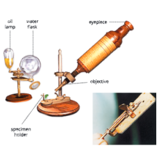
- Cells Were Discovered By Robert Hooke In 1665. The Invention Of Microscope Helped In The Discovery Of The Cell. Robert Hooke (1635-1703) Developed A Primitive Microscope By Using Two Lenses For Achieving Greater Magnification. In Such A Microscope, The Object To Be Seen Was Placed On A Stage Below And Light Coming From An Oil Flame Was Thrown On It By A Convex Mirror.
- He Observed Atliin Slice Ofcork (A Substance Obtained From The Bark Of A Tree) Under His Primitive Microscope. He Observed That The Cork Slice Had A Large Number Of Compartments Joined Together In A Honcycomb- Iike Pattern. He Named These Compartments As Cells (The Word Cell Comes From A Latin Word Celia Meaning A Compartment Or A Little Room). ’Ihis Was For The First Time That Anyone Had Observed That Living Things Consist Of Separate Units Called Cells.
- In 1674, Anton Von Leeuwenhoek (1632-1723) Made Further Improvements And Constructed A Simple Microscope That Could Magnify Up To 270 Times.
Fundamental Unit of Life Class 9 KSEEB notes
Cell – The Fundamental Unit Of Life
Let Us Perform The Following Activity To Find Out More About Cells.
Activity-1
- To Prepare Temporary Stained Mount Of Onion Peel To Study Epidermal Cells
You Will Need - Onion Bulb, Forceps, Watch Glass, Glass Slide, Camel Hair Brush, Covcrslip, Knife, Water, Razor Blade, Mounting Needle, Safranin Solution, Glycerine And Microscope.
Procedure
- Take An Onion Bulb, And Discard The Brown, Outer Dry Scales. With The Help Of A Knife, Cut The Onion Bulb Into Four Pieces. Remove One Fleshy Scale.
- Bend The Outer (Convex) Surface Of The Fleshy Scale Towards Yourself. Do Not Break It Completely So That The Two Halves Remain Attached By A Thin Transparent Strip Of Epidermal Peel.
- Gerilly Pull Llie Broken End. You Will Find Dial Die Thin Transparent Layer Of Epidermis Is Peeled Off Easily. Using A Pair Of Forceps, Remove The Peel And Place It On A Watch Glass Containing Water. ‘Ihis Will Prevent The Peel From Getting Folded Or Dry.
- With The Help Of A Blade, Cut A Square Piece Of This Peel And Place It In A Drop Of Water On A Clean Glass Slide With The Help Of A Fine Camel Hair Brush. Make Sure That The Peel Is Perfectly Flat On The Slide.
- Put A Drop Of Safeanin Solution On The Ped To Stain It And Leave It For 2-3 Minutes. Put A Drop Of Glycerine Solution Over It To Prevent It From Drying With The Help Of A Mounting Needle, Cover The Peel With A Coverslip, Taking Care That There Are No Folds Or Air Bubbles In It
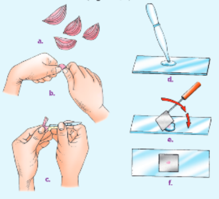
- Observe The Slide Under The Low Magnifying Power Of The Microscope. For Better Details, Increase The Magnification And Observe Under The High Power Of The Microscope.
Observations
- While Observing The Slide Under The Microscope, You Will Observe That The Epidermis Is One Cell Thick. These Structures Or Cells Look Similar To Each Other- Each Cell Has A Cell Wall, Cell Membrane, Nucleus, Cytoplasm And A Vacuole.
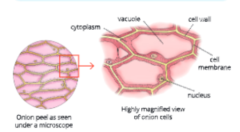
- The Cells That You Observed In The Above Activity Are The Basic Building Units Of The Onion Bulb. The Cells Of Onion Peel Are Rectangular Or Polygonal In Shape .All The Cells Are Firmly Bound Together. Like Onion, All Organisms Are Made Up Of Cells. Some Organisms Arc Made Up Of One Cell While Others Arc Made Up Of Many Cells.
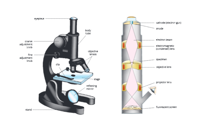
Microscope
- A Microscope Is An Instrument Which Is Used To Observe Objects That Are Invisible To The Naked Eye. For Example, Plant And Animal Cells, Bacteria, Fungi And Algae Can Be Observed Under A Microscope. Tire Two Common Types Of Microscopes Arc Compound Microscope And Electron Microscope.
SSLC Biology Chapter 1 The Fundamental Unit of Life
The Compound Microscope
- The Ordinary Light Or Compound Or Optical Microscope Used Extensively In Laboratories These Days Is A Greatly Improved Design Of Hooke’S Microscope It Consists Of Two Lenses, Namely, The Eyepiece Lens And The Objective Lens, Which Arc Combined To Produce A Greater Magnification. The Light Microscope Has A Magnification Up To 1500 Times, Good Enough To See Cells, Larger Organelles And Bacteria.
KSEEB Class 9 Biology Chapter 1 The Fundamental Unit Of Life Explained
Electron Microscope
- An Electron Microscope (Em) Has Much Greater Powers Of Magnification And Resolution Than Those Of An Optical (Light) Microscope. An Electron Microscope Can Resolve Points 1 Nm Apart. In This Microscope, A Beam Of Electrons Is Passed Through The Section Of A Material
To Produce The Image. The Electron Beam Passing Through The Specimen Section Is Focused By Electromagnets And Is Projected Onto A Fluorescent Screen For Direct View Or Onto A Photographic Plate For Permanent Recording. The Resulting Photograph Is Called An Electron Micrograph.
Electron Microscope Activity-2
- To Prepare Temporary Stained Mount Of Leaf Peels, Root Tips Of Onion And Peels Ol Onion Of Different Sizes And Observe Epidermal Cells.
You Will Need - Tradescantia Leaf, Onion Bulbs Of Different Sizes, Onion Roor Tips, Forceps, Watch Glass, Glass Slide, Water, Razor Blade, Iodine Solution And Microscope
Procedure
- For Preparing The Temporary Stained Mounts Of Peels Of Onion Bulbs Of Different Sizes, Follow The Steps Of Activity 1 As Given Before. Observe Different Sections Under A Microscope And Compare Their Structures.
- For Preparing The Temporary Stained Mount Of Leaf Peels, Tale The Leaf Of Tradtscaniia Plant. Take Out A Small Segment Of Peel From The Lower Surface Of The Leaf With A Jerk. Make T Temporary Mount Of This Peel As Mentioned In Activity 1 And Observe Under The Microscope.
- Similarly, You Can Prepare Temporary Stained Mount Of Onion Root Tips To Observe Epidermal Cells.
Observations
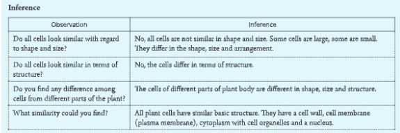
- What Do You Observe In The Above Activity? Do All Cells Look Similar With Regard To Shape And Size? Do All Cells Look Similar In Terms Of Structure? Do You Observe Any Difference Or Similarity Among Cells From Different Parts Of The Plant?
Activity-3
To Prepare A Temporary Mount Of Human Cheek Cells And Observe It Under A Microscope
You Will Need
A Clean Ice-Cream Spoon, Methylene Blue Solution, Glass Slides, Water, Needle And Microscope.
Procedure
Take An Icc-Crcam Spoon And Gently Scrape The Inner Lining Of Your Cheek. It Will Collect Some Viscous Transparent Material. With The Help Of A Clean Needle, Transfer This Material On A Clean Glass Slide.
Add A Drop Of Water To The Smear. Also Add A Drop Of Methylene Blue Solution To If. Leave The Preparation For About 1 Minute. Methylene Blue Is Used To Stain The Nucleus In A Cell Gently Place A Coverslip Over The Material With The Help Of A Needle To Avoid The Entry Of Air Bubbles. Press It Gendy In Between Folds Of A Rough Filter Paper To Remove Excess Fluid And For Uniform Distribution Of The Cells In The Mount Prepared.
Observe Under A Microscope And Find Out The Structural Details Of Cheek Cells.

Observation
What Do You observer What Is The Shape, Of The Ri*Fk You See? Draw It On The Observation Sheet.
Inference
- Under High Power Of A Compound Microscope, You Will Observe A Darkly Stained, Oval Or Spherical Dot-Like Structure Near The Centre Of The Cell.This Is The Nucleus. The Nucleus Is Surrounded By The Cytoplasm.
- The Cell Membrane Forms The Boundary Of A Cell Similar Structures (Nucleus) Were Also Found In The Onion Peel Cells. You Will Also Find That There Is No Large Central Vacuole Or Cell Wall As Observed In The Onion Peel (Plant) Cells.
- On The Basis Of Above Activities We Can Summarize That:
- A Cell Is The Structural And Functional Unit Of Living Beings.
- It Is Capable Of Independent Existence And Performs Essential Functions Of Life.
- A Cell Is Usually Microscopic (Ie. Invisible To The Naked Eye) In Nature.
- It Consists Of A Mass Of Protoplasm Surrounded By A Selectively Permeable Plasma Membrane.
- In An Animal Cell, The Plasma Membrane Is The Only Limiting Membrane. However, In Plant Cells, Fungi And Bacteria, The Cell Is Also Surrounded By A Cell Wall.
- All Organisms Start Their Life As A Single Cell. Every Cell Has Its Own Lifespan. The Old And Worn Out Cells Are Continuously Replaced By New Cells Except Few Cells Including Neurons.
- Many Cells Collectively Form A Tissue And Many Tissues Together Form An Organ. The Organs Work Together As A System For The Purpose Of Survival.
Class 9 Biology Karnataka Board Chapter 1 notes
Cell Theory
- The Cell Theory Was Proposed By German Botanist Matthias Schlcidcn (183$) And German Physician Theodore Schwann (1839). Later On In 1855, Rudolf Virchow, A German
- Pathologists added The Phrase Ottmis Cellula-E-Cdlula Meaning All Cells Arise From Pre-Existing Cells. The Major Points Of The Cell Theory Are As Follows:
- All Living Organisms Are Composed Of Cells.
- A Cell Is The Basic Structural And Functional Unit Of All Living Beings.
- All Cells Arise From Pre-Existing Cells.
Exceptions To Cell Theory
- Viruses Arc Non-Cellular Organisms. They Do Not Have Nucleus, Cytoplasm Or Enzyme And Do Not Perform Any Life Activity. They Can Multiply Only Inside The Living Host By Taking Over Their Machinery.
- Bacteria And Blue-Green Algae Are Not True Cells. They Do Not Have A Nuclear Membrane And Membrane Bound Cell Organelles.
Unicellular And Multicellular Organisms
- On The Basis Of The Number Of Cells, There Are Basically
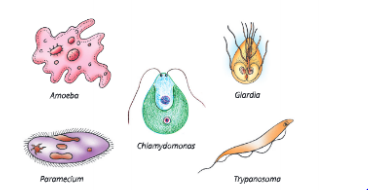
Two Types Of Organisms – Unicellular And Multicellular. - Unicellular (Uni Means Single) Organisms Are Made Up Of Only One Cell. For Example, Amoeba, Chlamydomonas, Euglena, Paramecium, Trypanosoma And Bacteria Are Unicellular Organisms.
- Multicellular (Multi Means Many) Organisms Are Made Up Of Many Cells That Group Together To Perform Many Functions Of The Body. For Example, Fungi, Plants And Animals Are Multicellular Organisms. All Multicellular Organisms Have Been Derived From A Single Cell, Zygote, Through Cell Division.
Prokaryotic And Eukaryotic Cells
On The Basis Of Their Nuclear Organization, Cells Have Been Classified Into Two Types:
• Prokaryotic Cells
• Eukaryotic Cells
Prokaryotic Cells(Gk. Pro: Before, Karyon: Nucleus)
- Prokaryotic Cells Are Single-Celled And Lack A Nuclear Membrane. These Cells Have Primitive Organization Of Genetic Material. The Genetic Material Is Equivalent To A Single Molecule Of A Circular Dna. These Cells Have An Undefined Nuclear Region Called The Nucleoid Due To Absence Of Nuclear Membrane. These Cells Lack Several Cytoplasmic Organelles Like Mitochondria, Lysosome, Endoplasmic Reticulum, Chloroplast And Nucleolus.
Many Of The Functions Of These Cells Are Performed By Poorly Organized Parts Of Cytoplasm. Bacteria And Blue-Green Algae Are Examples Of Prokaryotic Cells.
Eukaryotic Cells
(Gk. Eu: True, Karyotv Nucleus)
- Eukaryotic Cells Have A Well-Defined Nuclear Membrane. In These Cells, The Genetic Material Is Made Of Two Or More Linear Dna Molecules In The Nucleus, Enclosed In A Nuclear Membrane. These Cells Thus Have A Well- Organized Nucleus. These Cells Have Well-Developed Membrane-Bound Organelles, Such As Mitochondria, Endoplasmic Reticulum, Lysosome, Chloroplast And Nucleolus. Eukaryotic Cells Occur In Plants, Animals, Fungi And Protozoa.
The Fundamental Unit of Life KSEEB important questions
Cell – Shape, Size And Function
- Different Organisms Have Cells Of Different Kind. The Shape And Size Of Cells Are Related To The Specific Functions Performed By The Organisms.
Cell Shape
- Cells Show A Great Variation In Their Shapes (Fig. 1.8). Most Cells Have A Definite Shape. Cells May Be Spindle-Shaped – Muscle Cells, Elongated – Nerve Cells, Oval – Red Blood Corpuscles, Cuboidal – Germ Cells, Branched – Osteocytes And So On.
- Some Cells May Not Have Any Definite Shape, For Example, Amoeba And Leucocytes (White Blood 14 Corpuscles).

Class 9 SSLC Biology Chapter 1 textbook solutions
Cell Size
- The Smallest Known Cell Is Mycoplasma Or Pplo (Pleuropneumonia-Like Organism). Its Size Is 0.1 To 0. 5 Pm (Micrometre).
- The Bacterial Cell Is 0.5 To 5 Mm, Human Red Blood Corpuscles Are 7 To 20 Mm, Human Liver And Kidney Cells Arc 20 To 30 Mm And Nerve Cells Are About 90 To 100 Cm In Size.
Cell Function – Division Of Labour
- Each Cell Performs Certain Basic Functions That Are Characteristics Of All Living Beings. There Is A Division Of Labour In Multicellular Organisms And Also Within A Single Cell In Many Cases. This Means Different Parts Of The Body (Organs) Perform Different Functions. This Is Because Each Cell Has Certain Specific Components Within It Known As Cell Organelles. Each Cell Organelle Performs A Specific Function, Such As Producing Energy, Making New Materials (Proteins, Etc.), Clearing Up The Waste Material, Etc. These Organelles Together Constitute The Basic Unit, I.E. The Cell. A Cell Is Able To Perform Its Functions Because Of These Cell Organelles.
Structural Organization Of A Cell
- You Have Just Studied That Each Cell Has Special Components Called Cell Organelles. Although Cells Of Different Organisms Differ In Structure, Cells Within The Body Of A Multicellular Organism Also Differ In Shape, Size And Functions. In Spite Of These Differences, Every Cell Shows The Same Basic Structure – Cell Membrane Or Plasma Membrane, Nucleus And Cytoplasm. The Generalized Structures Of A Plant Cell And An Animal Cell Are Given
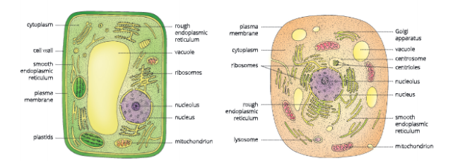
Cell Membrane Or Plasma Membrane
- Ever Cell Is Bound By A Thin Delicate Membrane Called Cell Membrane Or Plasma Membrane.
- Plasma Membrane Is The Outermost Covering Of The Cell Which Separates The Contents Of A Cell From Its External Environment.
We Can Sec Plasma Membrane Only With The Help Of An Electron Microscope. Structurally, Plasma Membrane Is Very Flexible. It Is Made Up Of Organic Molecules Called Lipid (Which Are Present In A Viscous Bilayer) And Protein (Within Lipid Bilayer). - The Flexibility Of Plasma Membrane Helps The Cell To Engulf In (Take In) Food And Other Substances From Its External Environment. This Process Of Engulfing Food And Other Material From The Outside Environment Is Known As Eudocytosis. Protozoans Like Amoeba Get Their Food Through endocytosis.

Functions
- Plasma Membrane Is Selectively Permeable. Therefore, It Allows Or Permits Die Entry And Exit Of Only Selected Substances. It Prevents The Movements Of Some Other Substances Across It
- Plasma Membrane Bounds The Semi-Fluid Content Of The Cells.
- It Protects Die Cell From Injury And Provides An Outer Boundary To The Cell.
- It Allows The Flow Of Materials And Information Between Different Organelles Within Die Cell As Well As Between One Cell And Another.
- It Has Carrier Proteins For Active Transport
- How Does Movement Of Substances Take Place In And Out Of Cells?
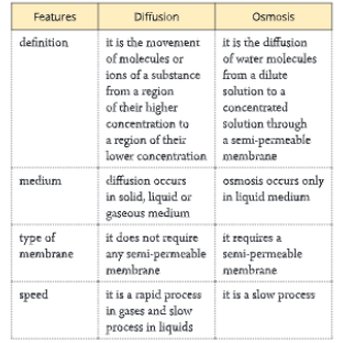
- The Movement Of Substances, In And Out Of The Cells, Occurs Through The Following Processes:
- Diffusion Osmosis
Diffusion
- The Movement Of The Molecules Of A Substance From Die Region Oftheir Higher Concentration To The Region Of Their Lower Concentration Is Called Diffusion.
- Importance Of Diffusion.
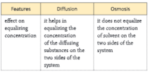
- Diffusion Plays An Important Role In Gaseous Exchange Between The Cells As Well As Between The Cell And Its Outside (External) Environment.
- It Is A Means Of The Movement Of Ions And Molecules Throughout Die Protoplast.
- Transpiration From Stomata Occurs By Diffusion.
- Aroma (Smell) Of Flowers Is Due To Diffusion Of Aromatic Compounds Of Flowers To Attract Pollinators.
- Carbon Dioxide Moves Out Of Die Cell Through Diffusion: There Are Many Metabolic Activities Like Respiration Taking Place Inside The Cell. During Cellular Respiration, Waste Like Carbon Dioxide Is Produced. This C02 Accumulates In The Cell In High Concentration. On The Other Hand, Concentration Of C02 Outside The Cell Is Lower Than That Inside The Cell. As A Result, C02 Moves From A Region Of Its High Concentration (Inside Die Cell) To A Region Of Low Concentration (Outside The Cell) Through The Process Of Diffusion. This Process Continues Till An Equilibrium (Even Distribution) Is Reached. The Diffusion Slows Down As Equilibrium Is Approached.
- Oxygen Moves Inside Die Cell Through Diffusion: Inside Die Cell, Oxygen Is Regularly Needed For Respiration And Other Metabolic Reactions. Thus, Die Concentration Of Oxygen Inside Die Cell Decreases. As A Result, Oxygen Moves From A Region Of Its High Concentration (Outside The Cell) To A Region Of Its Low Concentration (Inside The Cell).
Karnataka Board Class 9 Biology Chapter 1 summary
Osmosis
- The Diffusion Of Water Molecules Through A Scmi-Pcrmeable Membrane From A Region Where Water Is More Concentrated To A Region Where It Is Less Concentrated Is Called Osmosis.
- In Other Words, Osmosis Is The Diffusion Of Water From Its Pure State Or Dilute Solution Into A Stronger Or Concentrated Solution Through A Semi-Permeable Membrane.
Importance Of Osmosis
- Entry Of Soil Water Into Roots Occurs Through Osmosis.
- Cell To Cell Movement Of Water Occurs Through Osmosis.
- Living Cells Remain Turgid By Osmosis.
- The Stomata Open And Close In Response To Increase Or Decrease In Osmotic Pressure Of The Guard Cell.
Activity-4
- To Study Osmosis With The Help Of An Egg Yoa Will Need
A Hens Egg, Glass Beakers, Dilute Hydrochloric Acid, Salt
Solution And Water
Procedure
Take A Hen’S Egg And Remove Its Shell By Placing It In Dilute Hydrochloric Acid For Some Time. Since The Egg Shell Is Mosdy Calcium Carbonate, It Dissolves In Dilute Hcl
As The Egg Shell Is Dissolved, A Thin Outer Skin
Encloses The Egg. Put The Egg In Water For 5 Minutes And Observe.
What Do You Observe?
Observation 1
You Will Observe That The Egg Swells Because Water Passes Into It By Osmosis.
‘• Now, Take Another De-Shelled Egg And Place It In A Concentrated Salt Solution.
Again Observe For 5 Minutes. What Do You Observe?
Observation 2
You Will Observe That The Egg Shrinks. This Is Because Water Passes Out Of The Egg Into The Salt Solution As The Salt Solution Is More Concentrated.
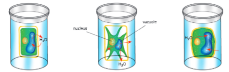
Isotonic, Hypotonic, And Hypertonic Solutions
- If You Take A Plant Cell And Place It In Solutions Having Different Concentrations, Then You Will Find That The Cell Shrinks In Hypertonic Solution, Swells In Hypotonic Solution While It Remains Unchanged In Isotonic Solution
If The Cell Is Kept In An Isotonic Solution
- Isotonic Solution Is One Which Has The Same Water Concentration As That In The Cell. In Such A Solution, There Is No Net Movement Of Water Across The Cell Membrane. In This Case, There Is A Movement Of Water Across The Cell Membrane In Both Directions. However, Tlie Amount Of Water Entering Die Cell Is Exaedy the Same As The Amount Of Water Leaving The Cell. As A Result, There Is No Overall Or Net Movement Of Water And Cell Retains The Same Size.
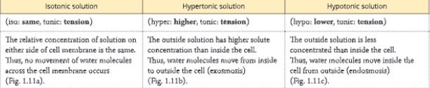
If The Cell Is Kept In A Hypertonic Solution
- In A Hypertonic Solution, The Concentration Of Water Molecules In The Outside Medium Is Lower Than That Inside The Cell. That Means The Solution Becomes Very Concentrated. Water Moves Through The Cell Membrane In Both Directions, But More Water Leaves The Cell Than Entering It As A Result, The Cell Shrinks.
If The Cell Is Kept In Hypotonic Solution
- In Hypotonic Solution, The Medium Surrounding The Cell Has Higher Water Concentration Than Inside The Cell. If You Place A Plant Cell In Hypotonic Solution, Then The Cell Will Swell Due To Osmosis. This Happens Because More Water Molecules From Outside Enter The Cell Than Leaving It. As A Result, The Cell Swells Up. Unicellular Organisms Living In Fresh Water And Most Plant Cells Absorb Water Through Osmosis.
- Roots Also Absorb Water Through Osmosis. This Happens Because The Air Spaces In Root Hair Contain Cell Sap Which Has High Concentration Of Salt And Less
Activity-5
To Study Osmosis With The Help Of Dried Raisins Or Apricots
You Will Need
Dried Raisins Or Apricots, Concentrated Sugar Or Salt
Solution, Wrater And Petri Dish
Procedure
Take Few Dried Raisins Or Apricots And Place Them In Plain Water.
leave Them For A Few Hours.
Observation 1
You Will Observe That Each Raisin Or Apricot Absorbs Water And Swells When Placed In Pure Water.
Now, Place These Raisins Or Apricots In Concentrated Sugar Or Salt Solution For Some Rime.
What Do You Observe?
Observation 2
You Twill Observe That The Raisins Or Apricots Lose Water And Then Shrink.
Concentration Of Water Than Outside. As A Result, Water Enters The Root Cell Through Osmosis.
Cell Wall
- In Addition To Plasma Membrane, Plant Cells Arc Surrounded By A Cell Wall Also. Cell Wall Is An Outer, Rigid, Protective And Supportive Covering Of Plant Cells. ‘Ihe Cell Wall Lies Outside The Plasma Membrane. Its Thickness Varies In Different Types Of Cells. The Cell Wall In Plant Cell Is Mainly Composed Of Cellulose. Cellulose Is A Complex Substance And Provides Mechanical And Structural Strength To Plants. Cell Wall Is Also Present In Fungi And Bacteria But Not In Animal Cell
Functions
- It Provides A Definite Shape To The Cell
- It Protects Plasma Membrane And Internal Structures From The Attack Of Pathogens And Mechanical Injury.
- It Counteracts The Osmotic Pressure.
- It Provides Rigidity To The Cell.
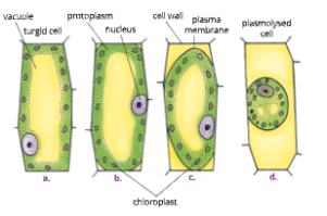
- A Living Plant Cell Can Lose Water Through Osmosis And As A Result There Is A Shrinkage Or Contraction Of Die Cell Contents Away From The Cell Wall. This Phenomenon Is Known As Plasmolysis. Let Us Perform The Following Activity To Learn More About Plasmolysis.
Activity-6
- To Show With The Help Of Rlioeo Leaf That Only Living Cells Undergo Plasmolysis
You Will Need - Rhoeo Leaf, Glass Slides, Concentrated Sugar Or Salt Solution, Wrater, Petri Dish And Microscope
- Procedure
- Take A Rhoeo Leaf And Break It To Take Out A Peel.
Mount This Peel Of Rhoeo Leaf In Water And Examine Under High Power Of Microscope. What Do You Observe?Observation 1 - You Will Observe Ihai The Leal’ Peel Contains Green Coloured Granules Called Chloroplnsts. The Chloroplasts Contain Chlorophyll, Which Helps In Photosynthesis.
Now, Put A Drop Of Strong Or Concentrated Solution Of Sugar Or Salt On The Leaf Peel. Wait For A Minute And Observe. What Do You Sec? - Observation 2
- You Will Observe That Plasmolysis Has Occurred And The Green Leaf Peel Cells Lose Water And Shrink Tire Cell Contents Away From The Cell Wall. This Is Because Water Leaves The Cells Through Osmosis.
Now, Place The Leaf Peel In Boiling Water For A Few Minutes. This Will Kill The Cells.
Mount This Peel Of Boiled Rhoeo Leaf In Water And Examine Under High Power Of Microscope.
Put A Drop Of Strong Or Concentrated Solution Of Sugar Or Salt Solution On The Leaf Peel. Wait For A Minute And Observe. What Do You See? Did Plasmolysis Occur This Time?
Observation 3
- You Will Observe That The Boiled Leaf Peel Did Not Show Plasmolysis. As The Plasma Membrane Is Dead, The Water Molecules Move Freely In And Out Of The Cell. This Shows That Only Living Cells Show Plasmolysis And Not The Dead Cells.
Plasmolysis
- Shrinkage Of The Protoplast (Cell Content) Of A Cell From Its Cell Wall Under The Influence Of A Hypertonic Solution Is Called Plasmolysis. If We Place A Living Turgid Cell In A Hypertonic Solution, Withdrawal Of Water (Exosmosis) Occurs From The Central Vacuole Of The Cell As A Result, Die Size Of The Protoplasm Becomes Reduced And The Plasma Membrane Is Withdrawn From The Cell Wall.
- Cells Of Plants Absorb So Much Water But Do Not Burst. Why?
- Did You Know That The Cells Of Plants, Fungi And Bacteria Can Withstand Very Dilute (Hypotonic) External Media Widiout Bursting? Hus Is Because Of Cell Wall. In Hypotonic Media, Cells Take Up Water Due To Osmosis. As A Result, The Cell Swells Building Up Pressure Against The Cell Wall. At The Same Time, Cell Wall Also Exerts Equal
Pressure Against The 3wou.Cn Cell Contents. Because Of
The Pressure Exerted By The Cell Wall, The Cells Of Plants, Fungi And Bacreria Can Withstand Much Greater Changes In The Surrounding Medium Than The Animal Cells.
Nucleus
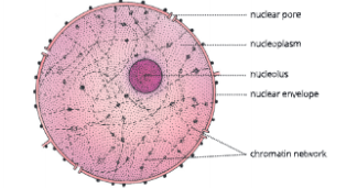
- A Dense, Generally Round (Spherical) But Sometimes Cylindrical Nucleus Is Present Almost At The Centre Of A Cell. The Nucleus Contains:
- Nuclear Envelope Or Nuclear Membrane,
- Nucleoplasm Or Nuclear Sap,
- Chromosomes (Chromatin Network), And Nucleolus
- Nuclear Envelope: It Is A Double-Membraned Structure, And Contains A Large Number Of Pores. It Separates The Nucleus From The Cytoplasm. The Nuclear Pores Control The
- Transfer Of Substances From Inside The Nucleus To Outside, I.E. To Cytoplasm.
- Nucleoplasm: Inside Die Nuclear Envelope Is Die Nucleoplasm. It Is Transparent, Semi-Fluid And Contains A Large Number Of Enzymes Which Are Required For The Syndvesis And Functioning Of Dna, Rna, Etc.
- Chromatin Network: In The Nucleus, Chromatin Network And Nucleolus Are Present. Chromatin Network (Gk. Chroma-Colour) Is A Tangled Fibrous Mass Of Thread-Like Structures.
- The Chromatin Threads
- Organize To Form Chromosomes Which Are Visible Only At The Time When The Cell Divides. Each Chromosome Consists Of Two Similar Threads Or Rod-Shaped Structures.
- Chromosomes Are Composed Of Dna (Deoxyribonucleic Acid) Molecules And Proteins. The Information For Inheritance Of Characteristics From Parents To The Next Generation
- Passes In The Form Of Dna. Dna Also Contains The Information For Cell Construction And Organization. Functional Segments Of Dna Are Called Genes. Genes Are The Carriers Of Heredity.
- Nucleolus: It Is A Dense, Round Structure Attached To A Chromatin Fibre At A Specific Region. The Nucleolus May Be One Or More In Number And Is Not Bound By Any Membrane. It Is Rich In Protein And Rna (Ribonucleic Acid) Molecules.
KSEEB SSLC Biology Fundamental Unit of Life Chapter 1 Revision Notes
Functions
- The Nucleus Controls Cell Metabolism And Other Activities Of The Cell, Hence, It Is Also Called Master Or Director Of The Cell.
- Chromatin Part Of The Nucleus Possesses All The Genetic Information That Is Required For The Growth And Development Of The Organism, Along With Its Reproduction, Metabolism And Behaviour.
- : Nucleus Plays A Central Role In Cellular Reproduction (Division Of Single Cell To Form Two Cells).
- Along With Controlling The Cell Environment, Nucleus Also Directs The Chemical Activities Of The Cell. This Determines The Development And Future Form Of The Cell.
Cytoplasm
- The Space Between The Plasma Membrane And The 20 Nucleus Is Filled By A Homogeneous, Translucent,
Colloidal Liquid Called Cytoplasm. It Consists Of Various Inorganic And Organic Molecules, Such As Water, Salts, Proteins And A Variety Of Enzymes.
Cytoplasm Also Contains Various Cell Organelles. These Cell Organelles Are Enclosed By Membranes. We Have Already Discussed That The Membrane-Bound Cell Organelles Are Present In Eukaryotes While They Are Absent In Prokaryotes
Functions
- The Cytoplasm Helps In The Intracellular (Within The Cell) Distribution Of Molecules, Enzymes And Nutrients.
- It Helps In The Exchange Of Materials Between Different Cell Organelles.
- Biosynthesis Of Nucleotides, Proteins And Fart)’ Acids Takes Place In The Cytoplasm.
Breaking Down Of Glucose Takes Place In The Cytoplasm.
Protoplasm
- Protoplasm Is The Living Component Of The Cell Containing Cytoplasm And The Nucleus In A Living Cell The Chemical Composition Of Protoplasm Varies From One Cell To Another.
- The Common Elements Found In Protoplasm Are Carbon, Hydrogen, Oxygen, Nitrogen, Iron, Phosphorus, Sulphur, Etc. Which Constitute Carbohydrates, Proteins, Fats, Minerals And Water. All The Living Components Of A Cell Lie In The Protoplasm And Perform Their Functions.
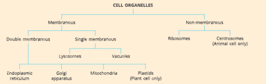
Cell Organelles
Each Cell Is Surrounded By A Cell Membrane. Cell Membrane Keeps The Contents Of The Cell Separate Front The External Environment. In Addition, Many Chemical Activities Take Place Continuously In Each Cell These Chemical Activities Are Required To Support The Complicated Structure And Functions Of Multicellular Organisms. Thus, To Perforin These Activities, The Cells Have Many Organelles That Float In The Cytoplasm. Some Organelles Are Membranous And Some Are Non- Membranous. Membranous Organelle Is A Characteristic
Feature Of Eukaryotes Only. Some Of These Organelles Are So Small That They Can Be Seen Only Under An Electron Microscope. Cell Organelles Carry Out Some Very Crucial Functions In Cells. The Different Cell Organelles That Are Found In The Cytoplasm Of The Cell Are Given
Endoplasmic Reticulum (Er)
- Endoplasmic Reticulum (Also Known As Er) Is An Interconnected Network Of Membrane-Lined Tubes And Sheets That Run Through The Cytoplasm. Ar Some Places It Is Connected With The Plasmalemma (Plasma Membrane) As Well As With Tire Nuclear Envelope. It Looks Like Round Or Oblong Bags Or Long Tubules. Endoplasmic Reticulum Is Similar In Structure To The Plasma Membrane. The Endoplasmic Reticulum Always Forms A Network System.
- Endoplasmic Reticulum Is Mainly Of Two Types – Smooth Er And Rough Er.
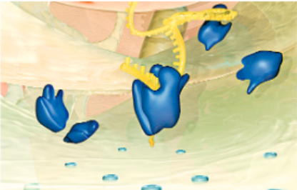
- Rough Endoplasmic Reticulum (Rer) Has A Rough Membrane Because A Number Of Ribosomes Are Found Attached To Its Outer
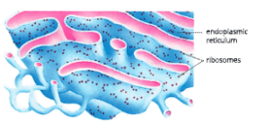
Surface. Ribosomes Are Present In All Active Cells. Ribosomes Are The Site For Protein Synthesis. Endoplasmic Reticulum Sends These Proteins To Different Sites In The Cell As Per Requirement. Rough Endoplasmic Reticulum Is Very Well-Developed In Plasma Cells, Fibroblasts, Goblet Cells, Etc.
Smooth Endoplasmic Reticulum (Ser) Does Not Have Any Ribosomes. This Type Of Endoplasmic Reticulum Is Found In Liver Cells, Interstitial Cells, Adipose Cells, Muscle Cells, Etc. It Helps In The Manufacture Of Fat Molecules And Lipids. Lipids Together With Some Proteins Help In The Building Up Of Cell Membrane Through A Process Known As Membrane Biogenesis. Many Other Proteins And Lipids Function As Enzymes And Hormones.
Class 9 KSEEB Biology Chapter 1 Cell Structure And Function Notes
Functions
- Endoplasmic Reticulum Divides The Cytoplasm Into Small Compartments. It Gives Rigidity To The Cell, Thus, Acts As A Skeletal Framework Of The Cell.
- It Serves As A Channel And Helps In Transporting Different Materials Such As Proteins Between Various Regions Of The Cytoplasm As Well As Between The Cytoplasm And The Nucleus. As It Helps In Intracellular Transport, It Is Also Known As The Circulatory System Of The Cell.
- It Acts As A Cytoplasmic Framework And Provides A Large Surface Area Inside The Cells For Various Biochemical Activities.
The Membranes Of Endoplasmic Reticulum Contain A Number Of Enzymes For Various Metabolic Activities. - In The Liver Cells Of Vertebrates, Smooth Endoplasmic Reticulum Helps In The Detoxification Of Many Poisons And Drugs.
Golgi Apparatus
Golgi Apparatus Was First Described By Camillo Golgi. It Is Present In All Eukaryotic Cells (Except Rbcs). Golgi Apparatus Consists Of A System Of Membrane- Bound Vesicles Which Are Arranged Somewhat Parallel To Each Other In Stacks Called Cisterns. These Membrane- Bound Vesicles Often Connect To The Membranes Of Endoplasmic Reticulum Forming Another Portion Of A 22 Complex Cellular Membrane System.
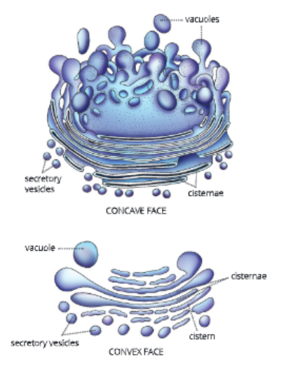
- There Arc Three Distinct Components Visible In The Golgi Apparatus, Flattened Sacs Or Cistemae, Clusters Of Tubules And Vesicles, And Large Vesicles Or Vacuoles.
- In Plant Cells, Golgi Apparatus Also Consists Of Many Freely Scattered Sub Units Called Dictyosomes.
The Fundamental Unit of Life Class 9 KSEEB PDF
Functions
- Golgi Apparatus Helps In The Secretion Of Mucus, Enzymes And Hormones. The Material Synthesized Near The Endoplasmic Reticulum Is Transported To Various Targets Inside
- And Outside The Cell Duough The Golgi Apparatus.
- It Helps In The Storage, Modification And Packaging Of Secretory Products In Die Vesicles.
- In Some Cases, Golgi Apparatus Also Helps In The Manufacture Of Complex Sugars From Simple Sugars. The Golgi Apparatus Also Helps In Die Formation Oflysosomes.
Ribosomes
Ribosomes Are Found In All Cells, Both Prokaryotes And Eukaryotes, Except In Mature Sperms And Rbcs. In Prokaryotic Cells, They Arc Found Floating Freely In
The Cytoplasm. In Eukaryotic Cells, They Occur Freely In The Cytoplasm As Well As Are Attached To The Outer Surface Of The Rough Endoplasmic Reticulum. They Are Also Found In The Mitochondria And Plastids.
Functions
Ribosomes Help In Protein Synthesis Inside The Cell Hence, They Arc Called Protein Factories Of The Cell.
Fundamental Unit Of Life KSEEB Chapter 1 Summary For Class 9
Lysosomes
- Lysosomes Are Membrane-Bound Sacs Filled With Digestive Enzymes. Ihese Enzymes Are Made By Rough Endoplasmic Reticulum. Lysosomes Have A Resistant Covering Membrane That Protects The Cell From The Digestive Enzymes Contained Inside Them.
- Lysosomes Help In Waste Disposal From The Cell. Lysosomes Digest Any Foreign Material As Well As Worn- Out Cell Organelles, And Hence, Keep The Cell Free From Any Unwanted Waste Material. Whenever Any Foreign Material (Such As Bacteria) Enters The Cell, The Lysosomes Break Them Up Into Small Pieces. This Is Because The Enzymes Released By The Lysosomes Are Very Powerful And Help In The Process Of Digestion Of These Foreign Particles, Hi Case The Cell Gets Damaged, Lysosomes Burst And Release The Enzymes, Which Digest Their Own Cell. Hence, They Are Called Suicide Bags Of The Cell.
Functions
- Lysosomes Help In Intracellular Digestion.
- They Provide Energy During Starvation By Controlled Breakdown Of Stored Food.
- Lysosomes Bring About Cellular Breakdown And Are Associated With Ageing.
Mitochondria – The Powerhouse Of The Cell
(Gk. Mitos: Thread, Chondrion: Granule)
Typically, The Mitochondria Are Sausage-Shaped Cell Organelles Covered By A Double-Membrane Envelope. The Outer Membrane Is Smooth And Porous, While The Inner Membrane Is Folded Into Structure Known As Cristae. These Folds Create A Large Surface Area For The Generation Of Atp (Adenosine Triphosphate) During Respiration. Mitochondria Release Energy In The Form Of Atp Molecules. This Energy Is Required For Performing Various Activities. Hence, Mitochondria Are Also Known As The Powerhouses Of Tire Cell Atp Is Known As The Energy Currency Of The Cell. Our Body Uses Energy Stored In Atp For Manufacturing New Chemical Compounds And For Mechanical Work.
The Cavity Of The Mitochondria Is Filled With Lipids, Proteins, Circular Dna And Ribosomes. Thus, Mitochondria Can Make Some Of Their Own Proteins.
Functions
Mitochondria Are Miniature Biochemical Factories Where Food Is Oxidized And Energy Is Released. This Energy Is Stored In The Form Of Atp. Hence, Mitochondria Are Called The Powerhouses Of The Cell.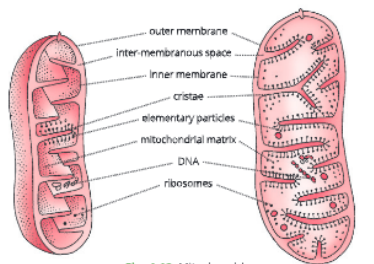 They Provide Important Intermediates For The Synthesis Of Several Biochemical Substances Like Chlorophyll, Cytochromes And Steroids.
Synthesis Of Many Amino Acids Occurs In Mitochondria.
Mitochondria Can Manufacture Some Of Their Own Proteins.
Plastids – Kitchen Of The Cell
Plastids Are Cell Organelles Found Only In A Plant Cell. Structurally, Plastids Contain Many Membrane Layers Embedded In A Material Called The Stroma. Like The Mitochondria, Plastids Also Have Their Own Dna And Ribosomes.
There Are Three Types Of Plastids – Chromo plasts (Coloured Plastids), Chloroplasts And Leucoplasts (White Or Colour less Plastids).
Chromo plasts (Gk. Chroma: Colour, Plasto: Formed) They Contain Fat Soluble Yellow, Orange Or Red Coloured Pigment. They Provide Colour To Flowers And Fruits. Chromoplasts Arc Formed Either From Leucoplasts Or Chloroplasts.
Chloroplasts (Gk. Chloros: Green,Plasto: Formed) They Are Green Plastids. Inside, Each Chloroplast Lias A Colourless Ground Matrix, Stroma And
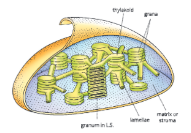
A Membranous System, Grana. Each Granum Has Membrane-Bound Sacs Called Thylakoids. Thylakoids Of Chloroplasts Possess Photosynthctic Green Pigment, I.E. Chlorophyll. They Also Contain Various Yellow Or Orange Pigments In Addition To Chlorophyll.
Leucoplasts (Gk. Leukos: White, Plasto: Formed) These Are Colourless Plastids And Are Named On The Basis Of The Substances They Store. They Store Starch, Oils And Protein Granules.
Functions
- Chromoplasts Arc Coloured Plastids That Provide Colour To The Flowers And Fruits.
- Chloroplasts, The Green Plastids, Help In Photosynthesis And Thus, Help In The Synthesis Of
- Food. These Are Called Kitchen Of The Cell.
- Leucoplasts Help In The Storage Of Food.
Vacuoles
- These Are Fluid-Tilled Membrane-Bound Spaces. Vacuoles Arc Storage Sacs For Liquid Or Solid Contents. They Are Bound By A Membrane Known As Tonoplast
- In Animal Cells, Vacuoles Are Small. In Mature Plant Cells, The Small Vacuoles Fuse To Form A Single Large Central Vacuole. Hie Central Vacuole Of Some Plant Cells May Occupy 50-90% Of The Cell Volume.
- In Plant Cells, Vacuoles Are Full Of Cell Sap And Provide Turgidity And Rigidity To The Cell. The Cell Sap Consists Of Tree Water And A Variety Ot Compounds In Solution Or Suspension. The Compounds Include Amino Adds, Minerals, Sugars, Organic Adds And Some Proteins.
- In Singlc-Ccllcd Organisms Such As Amoeba, The Vacuoles Are Modified As Food Vacuoles. The Food Vacuole Contains Food Items That Amoeba Iras Consumed. In Some Other Unicellular Organisms, Like Paramecium, These Vacuoles Are Spedalized To Expel Excess Water And Some Wastes From The Cell.
Functions
- Vacuoles Help The Plant Cells To Remain Turgid.
- They Play An Important Role In Growth By Helping In The Elongation Of Cells.
- They Provide An Aqueous Environment For Die Accumulation And Storage Of Water-Soluble Compounds (Sugars, Minerals, Pigments, Etc.).
- In Protozoans Like Amoeba And Paramecium, Vacuoles Help In Digestion And Excretion.
Centrosome
Centrosome Is A Small, Naked Protoplasmic Structure Present Near The Nucleus. It Is Present Only In Animal Cells. Centrosome Consists Of Two Small Granules Called Centrioles, Which Lie At Right Angles To Each Other. During Cell Division, Centrioles Migrate To Die Opposite Poles Of The Cell.
Functions
- They Help In Spindle Formation During Cell Division. They Act As Basal Bodies And Give Rise To Cilia And Flagella.
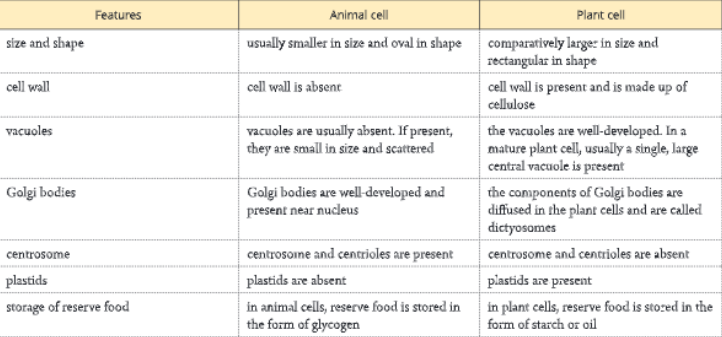
- Differences Between An Animal Cell And A Plant Cell
- The Differences Between An Animal Cell And A Plant Cell Are Given
Cell Division
- All New Cells Arise From Pre-Existing Cells By The Process Of Cell Division. New Cells Are Required For Growth, Replacement Of Old And Worn Out Cells And To Form Gametes During Reproduction. Cell Division Is Primarily Of Two Kinds Mitosis And Meiosis.
Mitosis
- Mitosis Or Mitotic Cell Division Is An Equational Division In Which One Parent Cell Divides To Form Two Daughter Cells. The Daughter Cells Are Identical To Each Other And Also To The Parent Cell In Every Respect.
- In Mitosis, The Same Normal Chromosome Number Of The Parent Cell Is Maintained At Each Stage Of Mitotic Division Of The Cell.
SSLC Class 9 Biology The Fundamental Unit of Life worksheet
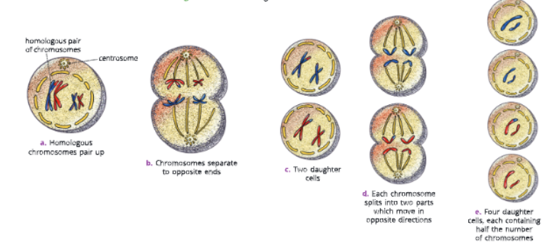
Meiosis
- Meiosis Takes Place In The Reproductive Cells That Produce Gametes, Sperms And Ova. Meiosis Is A Modified Mitosis In Which Chromosomes Divide Once And The Nucleus Divides Twice. As A Result Of Which The Number Of Chromosomes Is Reduced To Half. Hence Meiosis Is A Reductional Division.

Summary
- A cell is the structural and functional unit of life.
- The cell was discovered by Robert Hooke in 1665 while he was studying a thin slice of cork under a self-built microscope.
- The cell theory was formulated by two biologists, MJ Schlelden (1838) and T Schwann (1839).
- On the basis of nuclear organization, cells are of two types – prokaryotic cells and eukaryotic cells.
- A cell is enclosed by a plasma membrane which is made up of lipids and proteins. Plasma membrane is a living membrane. It is selectively permeable and allows only selected substances to pass through it.
- In plant cells, a cell wall is also present. It is mainly composed of cellulose and is located outside the cell membrane. Cell wall provides a definite shape to the cell. It protects plasma membrane and internal structures from pathogens and mechanical injury.
- In eukaryotes, the nucleus is separated from the cytoplasm by a double-layered membrane. It controls all metabolic and other activities of the cell. Hence, it is called the master of the cell.
Karnataka State Board Class 9 Biology Chapter 1 explanation
- Endoplasmic reticulum helps in intracellular transport. Hence, it is known as the circulatory system of the cell. It helps in the synthesis and transport of proteins and fats.
- Ribosomes help in protein synthesis inside the cell. Hence, they are called protein factories of the cell.
- Golgi apparatus consists of a system of membrane-bound vesicles which are arranged somewhat parallel to each other in stacks called cisterns.
- Golgi apparatus helps in the storage, modification and packaging of substances manufactured in cell.
- Mitochondria are miniature biochemical factories, where food is oxidized and energy is released. The energy is stored in the form of ATP. Plastids are cell organelles found only in plant cells.
- Plastids are of three types – chromoplasts, chloroplasts and leucoplasts. Chloroplasts help in photosynthesis, and thus in the synthesis of food. These are called kitchen of the cell. Lysosomes help in Intracellular digestion. They also bring about cellular breakdown, and hence are called suicide bags of the cell.
- In animal cells, vacuoles are usually absent. If present, they are small and scattered. But in mature plant cells, usually a single large central vacuole is present. It helps in maintaining the turgidity of the cell and stores important substances including waste.
Part A – Our PASTS – III (History)
- Chapter 1 How, When and Where
- Chapter 2 From Trade to Territory The Company Establishes Power
- Chapter 3 Ruling the Countryside
- Chapter 4 Tribals Dikus and the Vision of a Golden Age
- chapter 5 When People Rebel 1857 And After
- Chapter 6 Colonialism and the City: The Story of an Imperial Capital
- Chapter 7 Weavers, Iron Smelters and Factory Owners
- Chapter 8 Civilising the “Native” Educating the Nation
- Chapter 9 Women, Caste and Reform
- Chapter 10 The Changing World of Visual Arts
- Chapter 11 The Making of the National Movement 1870s -1947
- Chapter 12 India After Independence
Part B – Resources and Development (Geography)
- Chapter 1 Resources
- Chapter 2 Land,Soil, Water, Natural Vegetation and A wildlife Resources
- Chapter 3 Mineral and Power Resources
- Chapter 4 Agriculture
- Chapter 5 Industries
- Chapter 6 Human Resources
Part C: Social and Political Life -III (Civics)
- Chapter 1 The Indian Constitution
- Chapter 2 Understanding Secularism
- Chapter 3 Why Do We Need a Parliament ?
- Chapter 4 Understanding Laws
- Chapter 5 Judiciary
- Chapter 6 Understanding Our Criminal Justice System
- Chapter 7 Understanding Marignalisation
- Chapter 8 Confronting Marginalisation
- Chapter 9 Public Facilities
- Chapter 10 Law and Social Justice