Karnataka Board Class 11 Biology Histology And Anatomy Of Flowering Plants Synopsis
Internal Morphology deals with the study of the internal structure of different plant
organs. It has two branches they are Histology and Anatomy.
- Histology is the study of different tissues present in the plant body.
- Anatomy deals with the study of gross internal details of plant organs like roots, j stems, leaves, flowers, etc.
- Tissues are functional units of an organ. Tissues are groups of cells having similar, functions and origins.
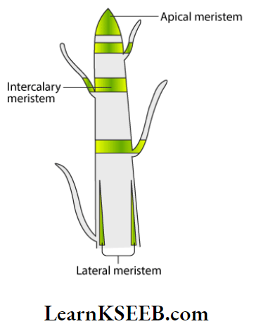
- Meristems are localised growth regions of the plant producing new tissues and organs throughout its life period. Based on origin- 2 types- primary meristem, and secondary meristem. Based on position – 3 types – apical, intercalary and lateral meristems.
Karnataka Board Class 11 Biology Anatomy of Flowering Plants notes
- Permanent tissues have mature cells adapted to specific functions. Simple tissues have similar kinds of cells.
- Parenchyma is a fundamental living tissue with intercellular spaces.
- Collenchyma is a simple living mechanical tissue. Sclerenchyma is a simple dead mechanical tissue with lignified walls.
- Tissue systems are three types. They are- epidermal, ground and vascular. The epidermal tissue systems are made up of epidermal cells, stomata arid the epidermal appendages.
- The ground tissue system forms the main bulk of the plant. It is divided into three zones- cortex, pericycle and pith.
- The vascular tissue system is formed by the xylem and phloem. The general plant internal structure is similar in both dicot and monocot roots.
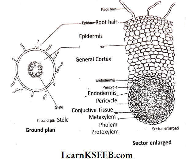
- Root TS shows 3 regions – epidermis, cortex and stele. The epidermis is an outermost layer with unicellular root hairs. Cuticle & stomata are absent. Exodermis contains suberised cells to prevent leakage of water.
- The general cortex is made up of parenchyma. Exodermis shows Casparian bands.
Stele consists of porlcyclo, vascular bundles, conjunctive tissues and pith. Pericycle is a singlo Inyorod pnronchymatoua cells. Poricyclo gives lateral roots. Vascular bundles are separate, radial, alternate and closed. Xylem i3 exarch. Stele is a monarch to autarch, generally tetrarch in dicot root and polyarch in monocot root.
- Pith is either scanty or absent in dicot root. It is large and well-developed in monocot root. Conjunctive tissue is present between the xylem and phloem strands.
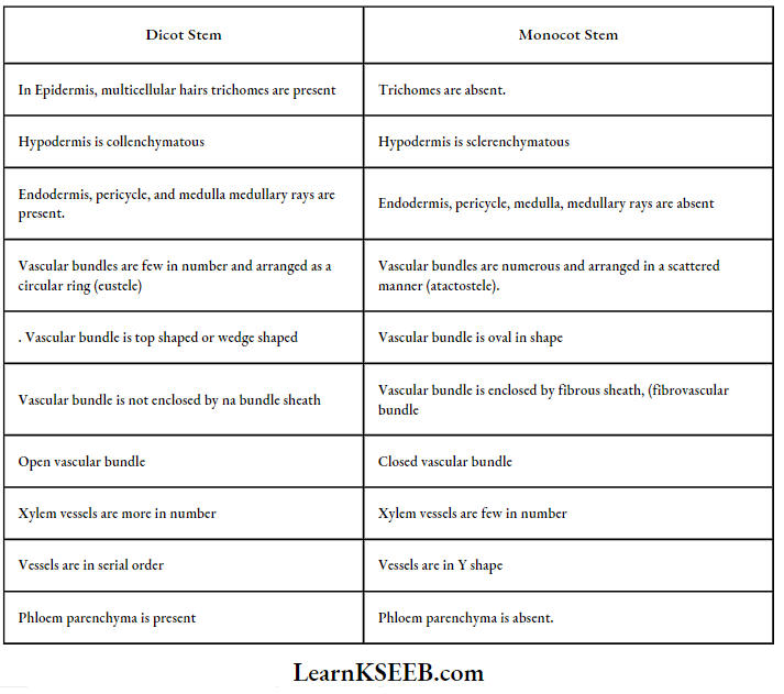
- A typical dicot stem consists of epidermis, cortex and stele. A typical monocot stem shows epidermis, hypodermis, ground tissue and vascular bundles. The epidermis is a unique protective layer covered with a cuticle. Stomata help in the exchange of gases.
- Hypodermis is coilenchymatous in the dicot stem and sclerenchymatous in the monocot I stem. The general cortex in the dicot stem and ground tissue in the monocot stem are parenchymatous.
- Endodermis is distinct only in the dicot stem with Casparian strips. Stele is eustele in dicot stem and atactostele in monocot stem.
Class 11 Karnataka Board Biology Chapter Anatomy of Flowering Plants Q&A
- Vascular bundles are few and arranged in a ring inside a pericycle in a dicot stem. They are numerous and scattered in the ground tissue in the monocot stem.
- Vascular bundles are conjoint, collateral and endarch. They are open in the dicot stem and closed in the monocot stem.
- Distinct pith and medullary rays are present in the dicot stem. The dicot leaf is dorsiventral and the monocot leaf is isobilateral.
- The epidermis covers the adaxial and abaxial surfaces of the leaf. Cuticle and stomata are present.
- Mesophyll is differentiated into upper palisade and lower spongy tissues in the dorsiventral leaf. It is undifferentiated arid homogeneous in an isobilateral leaf.
- Vascular bundles are collateral and closed. The xylem is on the upper side and the phloem is on the lower side. The vascular bundle is covered with a bundle sheath.
In Monocot leaf upper epidermis shows bulliform or motor cells. Secondary growth results in an increase in girth by the addition of secondary tissues. Interfascicular and intrafascicular cambia together form vascular cambium.
- The vascular cambium forms a secondary xylem inside and a secondary phloem on the exterior.
- Cork cambium develops from the cortical region. It produces cork towards the outside and secondary cortex on the inner side.
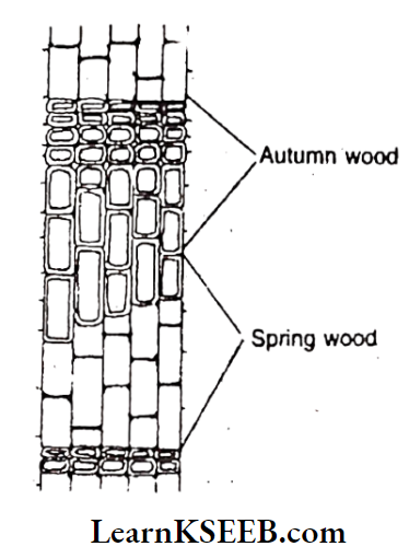
- Secondary xylem or wood forms the bulk of the tree. Production of secondary xylem is affected by seasonal change.
- Autumn wood and spring wood formed in one year together, constitute an annual ring. .
- Wood in the centre of the tree ceases to perform the function of conduction and is blocked by gums, resins, tyloses etc. It is called heartwood. Outer lighter sapwood is functional wood.
- Cork is a dead tissue containing suberised cells.
- Phellogen (cork cambium), phellem (cork) and phelloderm (secondary cortex) constitute periderm.
- Lenticels are lens-shaped openings found in the cork for gaseous exchange.
- All tissues outside the vascular cambium constitute bark.
Anatomy of Flowering Plants Very Short Answer Questions For Class 11
Question 1. The transverse section of a plant material shows the following anatomical features The vascular bundles are conjoint, scattered and surrounded by a sclerenchymatous bundle sheaths phloem parenchyma is absent. What will you identify it as?
Answer:
Monocotyledonous stem with closed vascular bundles.
Question 2. Why are the xylem and phloem called complex tissues?
Answer:
- Complex tissues are made of more than one type of cells that work together as a unit.
- Xylem and phloem are made of more than one type of cells i.e., parenchyma, fibres etc.
Question 3. How is the study of plant anatomy useful to us?
Answer:
Anatomy is useful to know the internal structure of the plant. It is useful in the classification of plants based on natural relations. It is useful to understand the plant functions, habitat of the plant and evolution of plants.
Anatomy of Flowering Plants Karnataka Board Class 11 important questions
Question 4. The protoxylem is the first formed xylem. If the protoxylem lies radially next to the phloem, what kind of arrangement of xylem would you call it? Where do you find it?
Answer:
- Radial arrangement
- They are found in roots.
Question 5. What is the function of phloem parenchyma?
Answer: Phloem parenchyma stores food materials and other substances like resins, latex and mucilage.
Question 6. What is present on the surface of the leaves which helps the plant to prevent loss of water but is absent in roots? What is the epidermal cell modification in plants which prevents water loss
Answer:
- Cuticle
- Bulliforni cells are an Isobilateral (monocotyledonous) leaf.
Question 7. Which part of the plant would show the following?
- Radial vascular bundle
- Polyarch xylem
- Well developed pith
- Exarch xylem
Answer:
- Radio vascular bundle – Root
- Polyarch xylem- Monocot root
- Well-developed pith – Monocot root
- Exarch xylem – Root
Question 8. What are the cells that make the leaves curl in plants during water stress? Give an example.
Answer:
- Large, colourless Bulliform cells
- Example: Monocot (Grass) leaves
Question 9. What constitutes the vascular cambial ring?
Answer:
- Intrafascicular cambium and interfasicular cambium.
- A cambial ring is formed in the dicot stem during secondary growth.
Question 10. Give one basic functional difference between phellogen and phelloderm.
Answer:
- Phellogen (cork cambium) is a meristematic tissue, formed from the primary cortex.
- Phelloderm (secondary cortex) is a permanent tissue formed by inner cells that are cut off from phellogen.
Question 11. If one debarks a tree, what parts of the plant are removed?
Answer:
- Periderm and secondary phloem are removed.
- All those tissues are exterior to the vascular cambium.
Question 12. Name the various kinds of dell layers which constitute the bark.
Answer: Periderm and secondary phloem.
Question 13. A student estimated the age of a tree to be about 300 years. How did he anatomically estimate the age of this tree ?
Answer: The age of a plant can be estimated by counting the number of annual rings.
Question 14. Assume that you have removed the duramen part of a tree. Will the tree survive of die?
Answer: Plant survives because of the presence of sapwood which is meant for the conduction of water and minerals. Duramen is not useful for conduction.
Karnataka Board Class 11 Biology Chapter On Flowering Plants Short Question And Answers
Question 1 . State the location and function of different types of Meristems.
Answer:
Based on location. Meristems are three types:
1. Apical meristems :
They are present at ” the growing tips of roots, stem, branches etc., They help in the linear growth of the plant body. These are primary meristems because they appear early in life and contribute to the formation of the primary plant body.
2. Intercalary meristem:
They are found in between permanent tissues. This meristem is separated from the. apical meristems during plant growth. They help in the linear growth of the stem and leaves. Growth of flowers and fruits after their initiation at the apex also occurs due to these meristems. They are active only for a short period. These [_ are also primary meristems.
Example: Meristems seen at the base of internodes and leaf bases of monocotyledons (particularly grasses).
3. Lateral meristems :
They are found on the lateral sides of the plant body. The cells divide periclinally and increase the thickness of the organs like the stem and root. These are secondary meristems.
Example: Vascular cambium that helps in secondary growth by producing secondary xylem and secondary phloem; phellogen (cork cambium) that helps in the formation of periderm.
Question 2. Cut a transverse section of the young stem of a plant from your garden and observe it under the microscope. How would you ascertain whether it is a monocot stem or a dicot stem? Give reasons
Answer:
Karnataka PUC 1 Biology Anatomy of Flowering Plants solved questions
Question 3. A transverse section of the trunk of a tree shows concentric rings which are known as annual rings. How are these rings formed? What is the significance of these rings?
Answer:
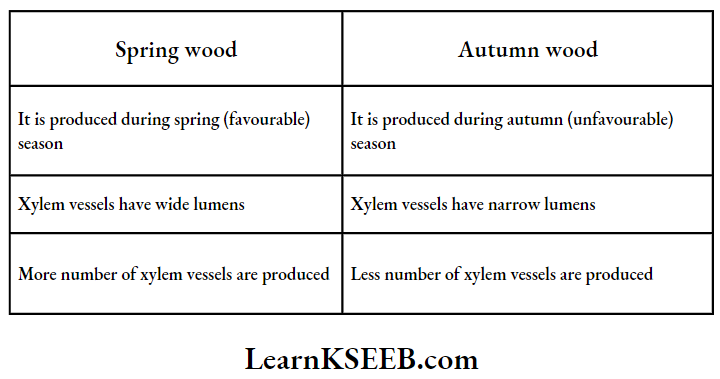
- In temperate and cold regions, the activity of cambium is influenced by seasonal variations.
- In favourable conditions, growth will be active. So plants require large amounts of water and minerals. During unfavourable conditions, plants are less active.
- In spring, the wood formed shows a greater number of xylem vessels having wide lumens. This is called spring wood or early wood.
- During autumn, wood formed shows less number of xylem vessels with narrow lumens. This is called autumn wood or latewood.
- Springwood and autumn wood appear alternately in the form of circles in the T.S. of a tree trunk. These are called Growth rings or annual rings.
- By counting the number of annual rings the age of a tree can be estimated approximately.
Secondary xylem in an annual ring:
Question 4. What is the difference between lenticels and stomata?
Answer:
Lenticels :
Lens-shaped openings in the cork of woody trees are called lenticels. They show closely arranged parenchymatous cells. The lenticels permit the exchange of gases between the outer atmosphere and the internal tissues of the woody organs. There is no opening and closing mechanism.
Stomata :
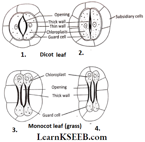
Stomata are present in the upper epidermis and lower epidermis of leaves. They help in the exchange of gases. In dicot leaves, on either side of stomata kidney-shaped guard cells are present. In monocot leaves, dumbbell-shaped guard cells are present. Guard cell contains chloroplast. They help in the opening and closing of the stomata. Stomata help in the gaseous exchange and also promote transpiration.
Question 5. Write the precise function of
- Sieve tube
- Interfasicular cambium
- Collenchyma
- Sclerenchyma
Answer:
- Sieve tube: The functions of the sieve tube are controlled by the nucleus of companion cells. The companion cells help in maintaining the pressure gradient in the sieve tubes. It transports food materials from leaves to other parts.
- Interfascicular cambium: The cells of medullary cells adjoining the intrafascicular cambium become meristematic and form interfascicular cambium. Thus a continuous ring of vascular cambium is formed.
- Collenchyma: The collenchyma cells which contain chloroplast are green in colour. Photosynthetic in function. Intercellular spaces are absent as the corners are thickened with pectin. So they provide tensile mechanical strength. It helps in the movement of the young stem, the petiole of the leaf, pedicle of the flower.
- Sclerenchyma: Sclerenchyma they are dead cells. Cell walls are thickened with lignin. Intercellular spaces are absent. So they, give mechanical strength to organs.
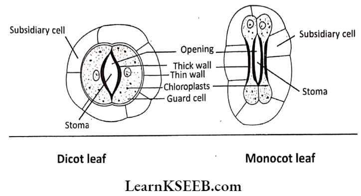
Question 6. The stomatal pore is guarded by two kidney-shaped guard cells. Name the epidermal cells surrounding the guard cells. How does a guard cell differ from an epidermal cell? Use a diagram to illustrate your answer.
Answer:
- The epidermal cells surrounding the guard cells are called subsidiary cells or accessory cells.
- The stoma is bounded by two kidney-shaped guard cells in dicots and dumbbell-shaped guard cells in monocots.
- Unlike that of other epidermal cells, guard cells possess chloroplast. The wall of the guard cells towards the stomatal pore is thick, while the outer wall is thin.
- The stoma, guard cell and subsidiary cells together constitute the stomatal complex.
Stomata of Dicot leaf & Monocot leaf:
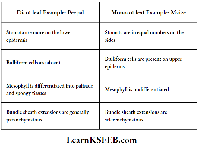
Question 7. Point out the differences in the anatomy of the leaf of peepal (Ficus religiosaj and maize (Zea mays].
Answer:
Dicot Leaf And Monocot Leaf:
Karnataka Board Class 11 Biology Chapter-wise question and answers
Question 8. Draw the diagrams and label the differences.
Answer:
1. The internal structure of the Dicot leaf:
Example: Peepal:
2. The internal structure of Monocot leaf:
Example: Maize
Question 9. The Cork cambium forms tissues that form the cork. Do you agree with this statement? Explain.
Answer:
Yes, Cork cambium or phellogen is a secondary meristematic tissue. It can divide. It divides and forms new cells on both sides. The tissue produced outside is called cork tissue or phellem. The tissue is produced towards an inner side in the secondary cortex or phelloderm.
Question 10. Name the three basic tissue systems in flowering plants. Give the tissue names under each system.
Answer:
The three basic tissue systems in flowering plants are
- Epidermal tissue system
- The ground or fundamental tissue system
- The vascular or conducting tissue system.
- The epidermal tissue system consists of parenchymatous tissue. They are epidermis, stomata and outgrowths.
- The ground or fundamental tissue system consists of simple tissues such as parenchyma, collenchyma and sclerenchyma.
- The vascular or conducting tissue system consists of complex tissues, the phloem and the xylem.
Question 11. Every 50 years, for 200 years, a nail was drilled into a tree to the same depth and at exactly 1m above the soil surface [assuming the ground level has not changed). What will be the pattern of the four nails on the tree? Do you know the reason for your answer? If yes give the reason.
Answer:
- The heads of all four nails are at the same level.
- The Stem of the plant undergoes later growth due to cambial activity. Hence, the growth (circumference) of the stem increases.
- All the four nails will be seen in the xylem portion of the stem.
- There will not be a change in nail position concerning vertical position from ground level. Because the vertical growth is reduced after some period and lateral growth is promoted in plants.
Question 12. Why is wood made of an xylem and not a phloem?
Answer:
- The cambial ring produces more xylem than phloem during secondary growth.
- Xylem except parenchyma, consists of dead tissues i.e., tracheids, vessels and fibres.
- Phloem is living complex tissue, except fibres (bast).
- Hence wood is made of xylem.
Long Answer Questions On Anatomy Of Flowering Plants for Class 11
Question 1. Explain the process of secondary growth in the stems of woody angiosperms with the help of schematic diagrams. What is its significance?
Or
What is periderm? How does periderm formation take place in the dicot stems?
Answer:
The growth of a stem or root in thickness due to the formation of secondary tissues due to the activity of primary and secondary meristems is called secondary growth.
Changes during the secondary growth of a dicot stem are divided into two groups.
They are
- Intrastelar secondary growth.
- Extrastelar secondary growth.
1. Intrastelar secondary growth :
The changes that occur inside the stele are called Intrastelar secondary growth. They are
Formation of vascular.cambial ring :
- Indicot stem, vascular bundles are in a circular ring. They are the open type with fascicular cambium.
- Parenchyma in medullary rays dedifferentiates into secondary meristem connecting fascicular cambium. These are called interfascicular cambiam.
- Fascicular and interfascicular cambia fuse to form vascular cambial ring.
The activity of vascular cambium :
- The vascular cambium has 2 types of initials.
- Fusiform initials: They give rise to the secondary xylem towards the centre and secondary phloem to the outside.
- Ray initials: They produce phloem rays towards the outside and xylem rays towards the inside.
- More secondary xylem is formed than secondary phloem.
- The secondary xylem is called wood and the secondary phloem bast.
- Stages of secondary growth in Dicot stem
- As the stem increases in thickness primary phloem and primary xylem are crushed and removed.
- The secondary xylem has vessels, fibres and xylem perenchyma. Vessels are pitted.
- The secondary phloem has sieve tubes, companion cells, fibres and phloem parenchyma.
- Xylem rays and phloem rays are also called vascular rays. They are helpful in lateral conduction and storage.
Stages of secondary growth in Dicot stem:
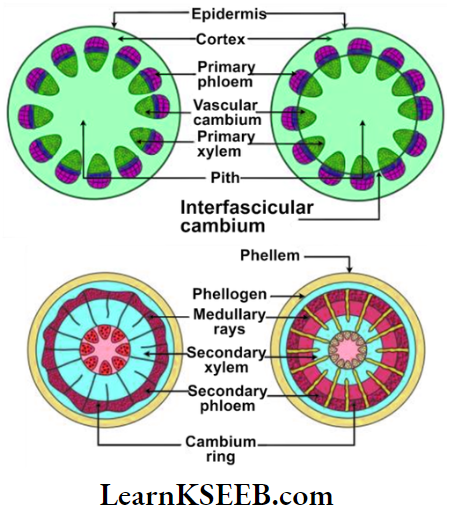
2. Extrastelar secondary growth: The changes which occur outside the stele are called Extrastelar secondary growth.
- Due to the formation of more secondary vascular tissues pressure is exerted on the epidermis causing its rupture. So a secondary protective layer (periderm) is formed.
- Parenchyma cells in the middle or inner cortex differentiate into a ring of secondary meristem. This is called cork cambium or phellogen. It cuts off new cells on both sides
- Tissue produced on the outside is called cork tissue or phellem. The tissue produced inside is called the secondary cortex or phelloderm.
- The phellogen, phellem, and phelloderm together constitute periderm.
- To facilitate gaseous exchange in the cork tissue certain bulged lens-shaped structures are formed. They are called lenticels.

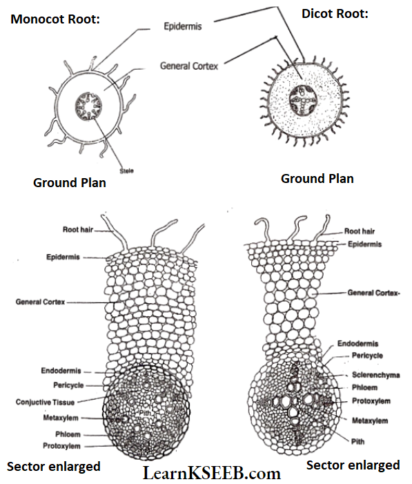
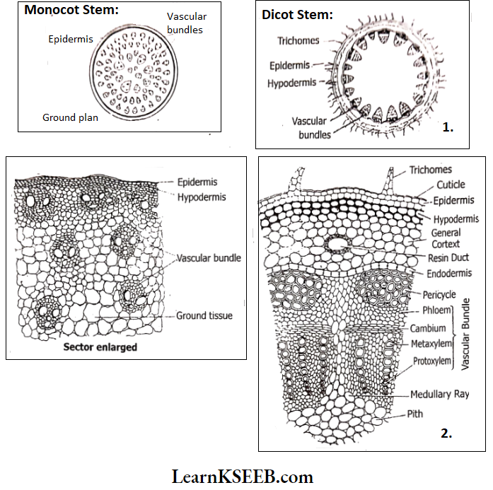
Question 2. Draw illustrations to bring out the anatomical differences between
- Monocot root and Dicot root
- Monocot stem and Dicot stem
Answer:
1. Monocot root and Dicot root:
2. Monocot stem and Dicot stem:
Class 11 Biology Karnataka Board Anatomy of Flowering Plants summary
Question 3. What are simple tissues? Describe various types of simple tissues.
Answer:
The tissues which are made of only one type of cells are called simple tissues. The various types of simple tissues are parenchyma, collenchyma and sclerenchyma.
Parenchyma :
- The Parenchyma is a living tissue. It occupies a major part of the plant body. So it is known as fundamental tissue or ground tissue.
- The cells are isodiametric. They may be spherical, oval-round, polygonal or elongated in shape.
- Cell walls are thin, and made up of cellulose.
- Intercellular spaces may be present or absent.
- Parenchyma performs functions like photosynthesis, storage, and secretion.
Collonchyma :
- Collenchyma Is a living mechanical tissue.
- It Is present below the epidermis in dicot plants.
- It is present as a continuous hypodermal ring
- Example: Helianthus annus or as a discontinuous ring (Cucurbita)
- Corners are thickened due to cellulose, hemicellulose and pectin.
- Intercellular spaces are absent.
- It provides mechanical support to the growing parts of the plant parts such as the young stem, petiole, pedicle etc.
- In the cytoplasm, if chloroplast is present, photosynthetic in function.
Scelerenchyma :
- Sclerenchyma is a dead mechanical tissue.
- Cells are long and narrow.
- Cell walls are thickened with lignin with pits.
- They are dead cells. Protoplast is absent.
- Based upon the structure, origin and development sclerenchyma are two types -fibres, and sclereids.
- Fibres are thick-walled, elongated and pointed cells.
- Sclereids (stone cells) are spherical, oval or cylindrical.
- Cell walls are highly thickened, lumen is very narrow.
- Sclereides are found in fleshy fruits like guava, pear and sapota.
- Seed coat of legumes, leaves of tea, fruit wall of nuts etc.
- Their main function is to give mechanical support.
Question 4. What are complex tissues? Describe various types of complex tissues.
Answer:
- Tissues which are made up of more than one type of cells and work together as a unit are called complex tissues.
- The xylem and phloem are complex tissues.
Xylem:
The main function of the xylem is to conduct water and minerals from the roots to the stem and leaves. Xylem also provides mechanical strength to plant parts
Xylem consists of:
- Tracheids
- Vessels
- Xylem fibres
- Xylem Parenchyma.
These are
- Tracheids: Tracheids are elongated or tube-like cells with thick and lignified walls and tapering ends. These are dead cells without protoplasm. Its main function is water transport
- Vessels: The presence of vessels is an important character found in angiosperms. Vessels
are absent in Gymnosperms. Vessels are dead cells without protoplasm. The cells are elongated or tube-like cells thickened with lignin. Its main function Is water transport. - Xylem fibres: These are highly thickened with lignin with a narrow central lumen. They may be septate or aseptate.
- Xylem Parenchyma: These are living cells. Cell walls are thickened with cellulose. They store food materials like starch, fats, and tannins. The ray parenchyma cell helps in the radial conduction of water.
Primary xylem is of two types – protoxylem and metaxylem.
The first formed primary xylem elements are called protoxylem. The later-formed primary xylem is called the metaxylem.
- In stems, the protoxylem is towards the centre and the metaxylem is towards the periphery. It is called endarch.
- In roots, the protoxylem is towards the periphery and the metaxylem is towards the centre. It is called an exarch.
Phloem:
The main function of phloem transport food materials usually from leaves to the other parts of the plant body.
Phloem contains:
- Sieve tube elements
- Companion cells
- Phloem parenchyma
- Phloem fibres.
These are
- Sieve tube elements: These are long, tube-like structures arranged longitudinally and are associated with companion cells. Their end walls are perforated in a sieve¬ manner to form sieve plates. A mature sieve tube element possesses a peripheral cytoplasm and a large vacuole but lacks a nucleus. The function of sieve tubes are controlled by the nucleus of companion cells.
- Companion cells: These are specialized parenchymatous cells which are closely associated with sieve tube elements. Both are connected by pit fields present between their common longitudinal walls.
- Phloem parenchyma: These are parenchyma cells in the phloem with tapering cylindrical cells which have dense cytoplasm and nucleus. They store food materials and other substances like resin, latex etc.
- Phloem fibres (bast fibres): These are sclerenchymatous cells. The cell wall is thick. At maturity, they lose their protoplasm and become dead.
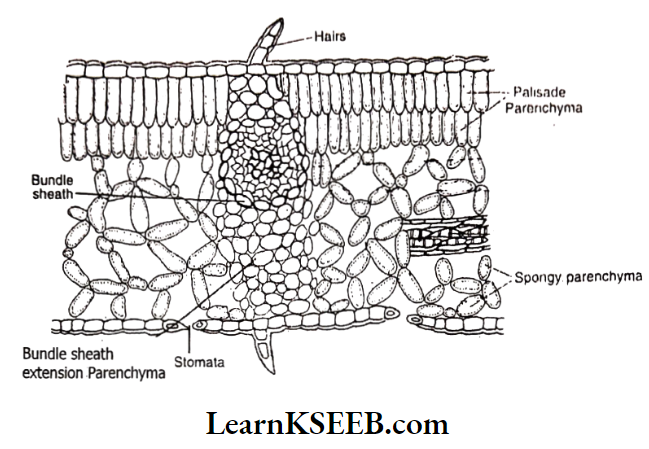
Question 5. Describe the internal structure of the dorsiventral leaf with the help of a labelled diagram.
Answer:
The transverse section of a dicot leaf or dorsiventral leaf shows three parts- epidermis, mesophyll and vascular bundles.
1. Epidermis :
- It is the outermost layer of leaf with one cell thickness.
- Cells are barrel-shaped. They are arranged compactly without intercellular spaces.
- The epidermis present on the upper (adaxial) side is called the upper epidermis.
- The epidermis on the lower (abaxial) side Is called lower epidermis
- The outer surface of the epidermis is covered by a waxy layer called a Cuticle.
- The epidermis shows multicellular hairs.
- Stomata are present. They are more on lower surface than the upper surface.
- The epidermis gives protection to the inner tissues. The cuticle regulates transpiration. Stomata help in exchange of gases.

2. Mesophyll :
Ground tissue present in between the two epidermal layers is called Mesophyll. It is Chlorenchymatous.
- In the dorsiventral leaf, mesophyll is differentiated into two parts- palisade parenchyma and spongy parenchyma.
Palisade parenchyma:
- Palisade parenchyma is found beneath the upper epidermis. Elongated and columnar cells are arranged in i – 3 rows.
- Intercellular spaces are narrow. Cells have a large number of chloroplasts nearer to the cell wall. (So the upper side of the leaf is dark green in colour). Palisade tissue is mainly concerned with the assimilation of carbohydrates.
Spongy parenchyma: Spongy parenchyma Is found towards the lower epidermis.
- It has 3 rows of irregularly shaped and loosely arranged cells. Intercellular spaces are large. Air cavities are found below the stomata.
- Cells have less number of chloroplasts. (So the lower side, of the leaf is pale green) Spongy Parenchyma facilitates gaseous exchange. They also help in the synthesis of food materials;
3. Vascular Bundle :
- Vascular bundles are extended in the mesophyll in the form of veins.
- Vascular bundles are bigger at the base of the leaf blade and gradually become smaller towards the margins and apex.
- Vascular bundles are conjoint, collateral and closed. Xylem is present towards the upper side and phloem towards the lower side.
- Vascular bundles help in the conduction of water, mineral salts and food materials.
- They also provide mechanical strength to the leaf.
- Each vascular bundle is enclosed by a layer of special mesophyll cells arranged compactly. This layer is called the Bundle sheath or Border parenchyma.
- Cells of the bundle sheath divide and extend towards both epidermal layers.
- These are called bundle sheath extensions. They help in the conduction of food materials from mesophyll cells to vascular bundles.
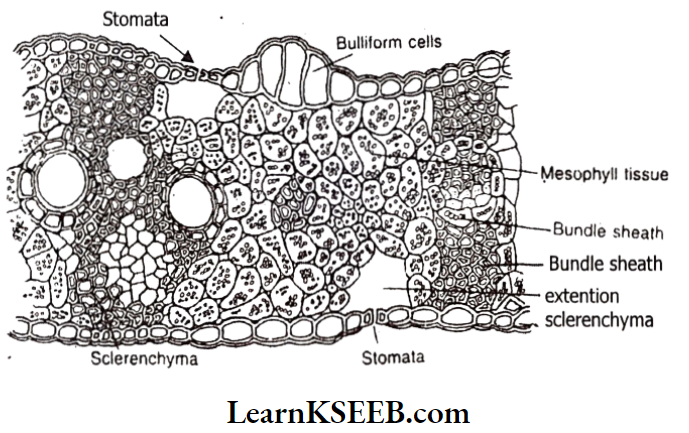
Question 6. Describe the internal structure of an isobilateral leaf with the help of a labelled diagram.
Answer:
- The transverse section of the monocot or isobilateral leaf shows three parts- epidermis, mesophyll and vascular bundles.
- This Is the outermost layer on both sides of the leaf. Cells are one cell in thickness. They are barrel-shaped and closely packed without Intercellular spaces.
- It is covered by a waxy layer called the cuticle.
- The epidermis on the adaxial (upper) surface is called the upper epidermis. The epidermis on the abaxial (lower) surface is called the lower epidermis.
- Hairs are absent. Stomata are present on both sides in equal numbers.
- In grasses, specialised cells are present in the upper epidermis. They are called bulliform cells or motor cells. They are thin-walled and filled with water.
- They help in rolling and unrolling of the leaf.
- The epidermis gives protection to inner tissues. The cuticle regulates transpiration.
- Stomata help in the exchange of gases.
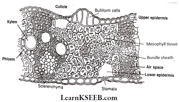
2. Mesophyll :
- Ground tissue present between two epidermal layers is called mesophyll.
- It is chlorenchymatous.
- Mesophyll is undifferentiated.
- Cells have chloroplasts and perform the assimilation of carbohydrates.
- Sometimes patches of sclerenchyma are found beneath the epidermis.
- They provide mechanical strength.
3. Vascular Bundles :
- Vascular bundles are present in the mesophyll in the form of veins.
- Vascular bundles are conjoint, collateral and endarch.
- Xylem is present on the upper side and phloem is present on the lower
side. - Veins help in the conduction of water, mineral salts and food materials. They
also provide mechanical strength. - Each vascular bundle is enclosed by a layer of special mesophyll cells called
bundle sheath or border parenchyma. - Cells present on either side of vascular bundles towards the upper and lower epidermis are Called bundle sheath extensions.
- In many monocots, they are sclerenchymatous and provide mechanical support.
Question 7. Distinguish between the following :
Answer:
- Exarch and endarch condition of protoxylem
- Stole and vascular bundle.
- Protoxylem and metaxylem
- Interfasicular cambium and intrafasicular cambium
- Open and closed vascular bundles
- Stem hair and root hair
- Heartwood and sap wood.
- Springwood and autumn wood.
Answer:
1. In roots, the protoxylem lies towards the periphery and the metaxylem lies towards the centre. Such type of protoxylem is called Exarch. In stems, the protoxylem lies towards the centre (pitch) and the metaxylem lies towards the periphery of the organ. Such a type of protoxylem is called Endarch.
2. Stele :
The stele is the central conducting cylinder. Generally it may have pericycle, vascular bundle, medulla and conjunctive tissue or medullary rays.
- Vascular bundles: Xylem and phloem are present in vascular bundles. Xylem conducts water and phloem conducts food materials.
3. Protoxylem: The first formed primary xylem elements are called protoxylem.
- Metaxylem: Later-formed primary xylem elements are called metaxylem.
4. Intrafasicular cambium:
Cambium present between the primary xylem and primary phloem is called Intrafasicular cambium. It is present inside vascular bundle (Intra = Inside; fascicular = vascular bundle)
- Interfascicular cambium : The cells in medullary rays become meristematic and form interfascicular cambium (Inter = in between; fascicular = vascular bundle)
5. Open vascular bundle: The vascular bundle which have a cambium between the xylem and phloem are called the open vascular bundle.
- Closed vascular bundle: In these vascular bundles cambium is absent between the xylem and phloem.
6. Stem hair and root hair:
- Stem hair : Multicellular hairs present on the stem an stem hair or trichomes. Their main function is to prevent the entry of pathogens.
- Root hair: Unicellular hairs present on the root are root hair. Their main function is absorption of water.
7. Heartwood and sapwood :
- Heartwood: The dark brown-coloured central part of the secondary xylem comprising of dead elements with highly lignified walls is called heart wood. It is infiltrated with various organic compounds like tannins, resins, oils, gums, aromatic substances and essential oils. The heartwood does not conduct water but it gives mechanical support to the stem.
- Sapwood: The peripheral region of the secondary xylem is lighter in colour and is known as sap wood. It conducts water and minerals from root to leaf.
8. Springwood and Autumn wood:
Question 8. What is stomatal apparatus? Describe the structure of stomata with a labelled diagram.
Answer:
Structure of stomata :
- Tiny pores in the epidermis of young aerial parts of the plant are called stomata.
- Stomata are more abundant in the leaf epidermis.
- The stoma is bounded by two kidney-shaped guard cells in dicots and dumbbell¬ shaped guard cells in monocots.
- Example: Grasses).
- Unlike that of other epidermal cells, guard cells possess chloroplasts.
- The wall of the guard cell towards the stomatal pore is thick, while the outer wall is thin.
- Epidermal cells surrounding guard cells are called subsidiary or accessory cells.
- They differ from other epidermal cells in their shape and position.
- The stoma is followed by an air cavity called a substomatal cavity in the mesophyll.
- The stoma, guard cells and subsidiary cells together constitute the stomatal apparatus.
Question 9. Describe the T.S of a dicot stem
Answer:
Internal structure of young dlcot stem – Example: Hellanthus annuus
The transverse section of a dicot stem shows three distinct zones – epidermis; cortex and stele.
1. Epidermis :
- The outermost layer in the young dicot stem is called the epidermis. It is one cell in thickness.
- Cells are tubular or rectangular. They are arranged compactly without intercellular spaces.
- The outer surface of the epidermis is covered by a waxy substance called cutin. This layer is called the cuticle.
- Minute pores found in the epidermis are called stomata.
- Multicellular. hairs developing on the epidermis are called trichomes.
- The epidermis gives protection to the inner tissues.
- Stomata facilitate the exchange of gases and promote transpiration.
- Cuticle and trichomes check transpiration. They also protect the stem from high temperatures.
- Trichomes also help in preventing the entry of pathogenic micro-organisms.
2. Cortex :
It is extrastelar ground tissue. It shows three subzones -hypodermis, general cortex and endodermis.
Hypodermis :
- The layer present below the epidermis is called hypodermis.
- It consists of 3- 6 layers of Collenchyma.
- Cells are arranged compactly without intercellular spaces. They show excessively thickened corners.
- Hypodermis helps in providing tensile strength to the stem.
- Hypodermis also helps in the production offood materials by having chloroplasts.
General Cortex :
- It is found below the hypodermis.
- It consists of 5-10 rows of parenchyma.
- Cells are isodiametric or oval or spherical.
- Resin or latex ducts may be present in it.
- Outer layers of cells have chloroplasts and perform assimilation of food materials.
- The inner layers are concerned with the storage of food.
Endodermis:
- It Is the Innermost layer of the cortex.
- It Is in one layer with barrel-shaped, compactly arranged cells.
- Radial and transverse walls show Casparian bands.
- Endodermal cells store starch grains. So it is known as a starch sheath.
3. Stele: A central conducting cylinder is called a stele. It occupies a major portion of the stem. It shows four parts.
Pericycle :
- It is the outermost layer of the stele.
- It lies, between endodermis and vascular bundles.
- It has alternate patches of sclerenchyma and parenchyma.
Vascular Bundles :
- Each vascular bundle is wedge or top-shaped.
- A limited number of vascular bundles are arranged in the shape of a circular ring. Such an arrangement is called Eustele.
- In each vascular bundle, the phloem is present outside and the xylem towards the inside on the same radius. So vascular bundle is conjoint and collateral.
- Meristematic tissue is present in between the xylem and phloem. It is called fascicular cambium. A vascular bundle with cambium is called an open type.
- The xylem is endarch (protoxylem towards the centre).
- Xylem has vessels and xylem parenchyma. Tracheids and fibres are also present. Xylem conducts water and salts.
- Phloem has sieve tubes, companion cells, phloem parenchyma and fibres. It conducts food materials.
Medulla :
The central part of the stele is called the medulla. It is filled with parenchyma. It is well-developed and extensive. It stores food materials.
Medullary rays :
- The medulla extends, to the periphery in between the vascular bundles forming medullary rays. Parenchymatous cells are living, thin-walled and elongate radially.
- Medullary rays connect the stele and cortex. They help in lateral conduction.
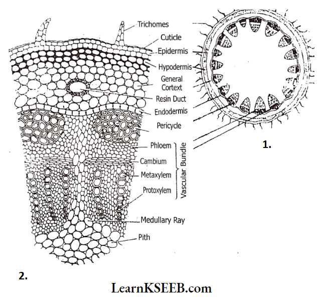
Question 10. Describe the T.S. of monocot stem.
Answer:
The internal structure of Monocot Stem :
The anatomy of the Monocot stem shows four distinct parts- Epidermis, hypodermis, ground tissue, and vascular bundles. A distinct cortex is absent. Endodermis, pericycle, medulla, and medullary rays are absent.
1. Epidermis
- The outermost layer is called the epidermis. It Is made up of living, rectangular or tabular cells. They are arranged compactly without Intercellular spaces.
- A waxy layer is deposited on the outer surface of the epidermis. This is called the cuticle.
- Trichomes are absent. Numerous stomata are found in the epidermis.
- The epidermis gives protection to inner tissues. Stomata help in the exchange of gases. Cuticle prevents evaporation of water.
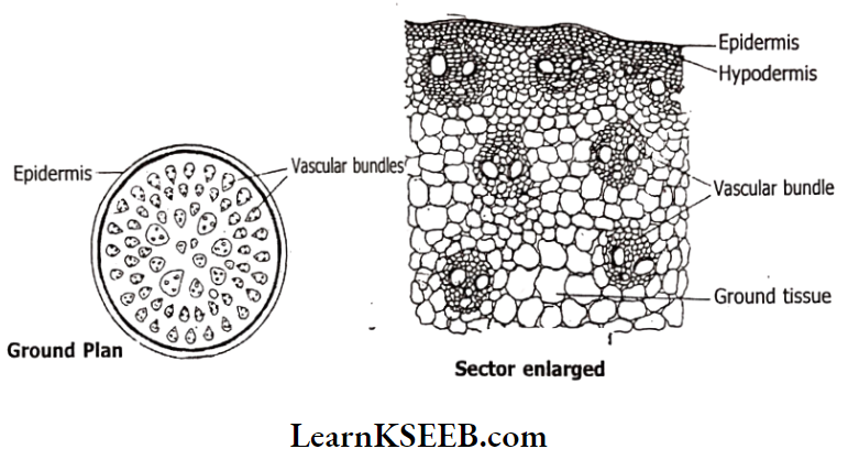
Hypodermis :
It is present beneath the epidermis. It is made up of 1- 4 rows of thick-walled sclerenchymatous fibres. Intercellular spaces are absent. It gives mechanical strength to the stem.
3. Ground Tissue :
- The tissue next to the hypodermis filling the remaining part of the stem (except vascular bundles) is called ground tissue.
- It is parenchymatous.
- Cells are thin-walled with or without chloroplasts. They are loosely packed with intercellular spaces.
- It is mainly concerned with the synthesis and storage of food materials.
4. Vascular Bundles :
- Numerous bundles are irregularly scattered in the ground tissue. Such an arrangement is called Atactostele.
- Inner bundles are bigger. Peripheral bundles are small in size. They are oval in shape.
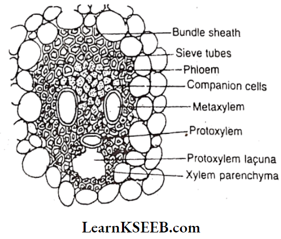
- Each vascular bundle is enclosed by a sclerenchymatous sheath. So it is called a fibrovascular bundle.
- Each bundle has a phloem towards the outside and a xylem towards the inside of the radius. So it Is described as conjoint and collateral. bundle on the same concerned with the conduction of water, salts and food
- Vascular bundles are materials.
- Cambium Is absent between the xylem and phloem. So the vascular bundle is a closed type.
- The xylem is endarch (protoxylem towards the centre). Xylem consists of tracheids, vessels, fibres and xylem parenchyma.
- The xylem is arranged in the shape of Y. Out of four xylem vessels, two are metaxylem and two are protoxylem vessels. It stores water.
- Phloem has sieve tubes and companion cells. Phloem parenchyma is an absent sheath
Karnataka Class 11 Biology handwritten notes Anatomy of Flowering Plants
Question 11. Describe the internal structure of a dicot root.
Answer:
The internal structure of the primary dicot root has three zones- epidermis, cortex, and stele. The cortex is bigger than the stele.
1. Epidermis :
- It is the outermost layer of thin-walled rectangular living cells arranged compactly without intercellular spaces.
- Cuticle and stomata are absent.
- Some epidermal cells (trichoblasts) produce tubular extensions called root hairs. Cells giving rise to root hairs are smaller than other epidermal cells. The Epidermis of the root is called rhizodermis or piliferous layer or epiblema due to the presence of root hairs.
- Root hairs absorb capillary water. The epidermis gives protection to the inner tissues.
2. Cortex :
The ground tissue system extending from the epidermis to the stele is called the cortex. It is differentiated into three parts.
Exodermis :
- The outermost layer of the cortex with 2 or 3 rows of suberised thick-walled cells is called exodermis.
- It acts as a protective layer when the epidermis is removed.
- It prevents the exit of water from the cortex.
General cortex :
- It is present beneath the exodermis.
- It has several layers of loosely arranged thin-walled parenchyma intercellular.
- spaces are present.
- Cells store food materials.
- The general cortex helps in the lateral conduction of water from the epidermis to the xylem vessels.
Endodermis :
- It is the innermost layer of the cortex.
- It is made up of a single layer of barrel-shaped cells.
- Radial and transverse walls of endodermal cells show thickening due to the deposition of lignin and suberin. These are called Casparian thickenings. It is a characteristic feature of the endodermis.
- Endodermal cells present opposite to protoxylem are thin-walled without Casparian strips. These cells are called passage cells.
- Passage cells help in the entry of water and salts from the cortex into the stele.
3. Stele: The central conducting cylinder Is called the stele. It shows three parts.
- Pericyte :
- It Is the outer layer of the stele. It Is uniseriate with thin-walled rectangular parenchymatous cells.
- Pericycle gives rise to lateral roots.
- It also helps in secondary growth.
- Vascular bundles: Xylem and phloem are arranged alternately on separate radii.
- So vascular bundles are separate or radial.
- Xylem and phloem conduct water and food materials respectively.
- The protoxylem is towards the pericycle and the metaxylem is towards the centre. So the xylem is exarch.
- Xylem is variable from monarch to octarch (xylem groups 1-8) usually tetrarch (4 xylem groups alternating with 4 phloem bundles). Monarch Trapa, Tetrarch – Gossypium, Octarch – Costanea,
- Cambium is absent.
- Parenchyma tissue extending between the xylem and phloem strands is called conjunctive tissue. It helps in the storage of food materials.
- Pith or Medulla:
- The central portion of the stele is called the medulla or pith. It may be completely absent in the dicot root.
- When it is present, it is parenchymatous. It helps in the storage of food and water.
Karnataka Board Class 11 Biology exam questions Anatomy of Flowering Plants
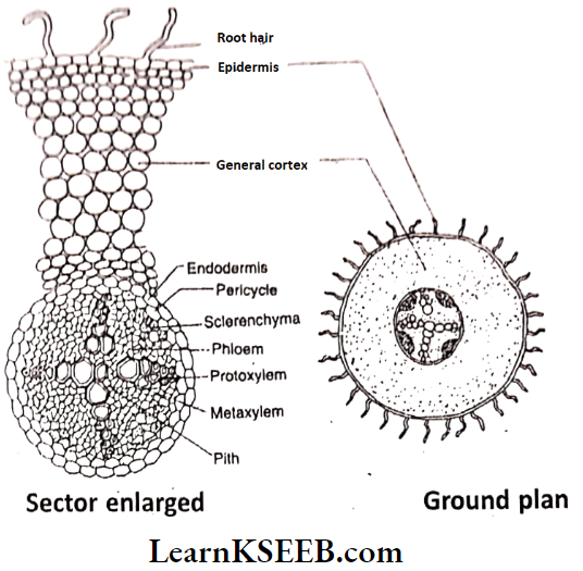
Question 12. Describe the internal structure of a monocot root.
Answer:
The internal structure of the monocot root is differentiated into three zones- epidermis, cortex and stele.
1. Epidermis :
- The outermost layer is called the epidermis. Cells are living, rectangular, thin-walled. They are compactly arranged without intercellular spaces.
- Cuticle and stomata are absent.
- Some cells (trichoblasts) show tubular extensions- root hairs. The Epidermis of the root having root hairs is called epiblema or piliferous layer or rhizodermis.
- Root hairs help in the absorption of capillary water from the soil. The epidermis gives protection to the inner tissues.
2. Cortex :
This is the ground tissue system extending from the epidermis to the stele. It is differentiated into three parts.
Exodermis :
- It is the outermost layer of the cortex with 2 or 3 rows of cells. Cell walls are thick and suberised.
- It acts as a protective layer when the epidermis is removed.
- It prevents the exit of water from the cortex.
General cortex :
- It is present beneath the exodermis.
- It has several layers of loosely arranged thin-walled parenchyma.
- Intercellular spaces are present.
- Cells store food materials lateral conduction of water Irom epidermis to xylem vessels.
Endodermis :
- It is the innermost layer of the cortex.
- It is made up of a single layer of barrel-shaped cells.
- Radial and transverse walls of endodermal cells show thickening due to the deposition of lignin and suberin. These are called Casparian thickenings.
- Endodermal cells present opposite to protoxylem are thin-walled without Casparian strips. These cells are called passage cells.
- Passage cells help in the entry of water and salts from the cortex into the stele.
3. Stele:
The central conducting cylinder is called a stele. It is very prominent and bigger in size. It shows three parts.
Pericycle :
- It is the outermost layer of the stele. It is. uniseriate with thin-walled parenchymatous cells.
- Pericycle gives rise to lateral roots.
- In old roots it becomes sclerenchymatous and gives mechanical strength.
Vascular bundles :
- The xylem and phloem are arranged alternately on separate radii. So vascular bundles are radial or separate.
- The protoxylem is towards the pericycle and metaxylem towards the centre. So xyl is exarch.
- The xylem is polyarch (numerous xylem groups).
- Cambium is absent.
- The Xylem Is concerned with the conduction of water and salts. Phloem conducts organic solutes.
- Parenchyma tissue extending between the xylem and phloem strands is called conjunctive tissue.
- Cells are rarely thick-walled. It is helpful in the storage of food and provides mechanical strength. mechanical strength.