Karnataka Board Class 11 Biology Biomolecules Synopsis
Even though there is a wide diversity in living organisms, all living organisms are made up of the same chemicals.
The elemental composition of living and non-living matter appears similar when analyzed qualitatively.
- Analysis reveals that the relative abundance of carbon and hydrogen concerning other elements is higher in any living organism than in the earth’s crust.
- The most abundant chemical in living organisms is water.
- All the carbon compounds that we get from living tissues can be called “biomolecules”.
- There are thousands of carbon compounds that we get from living tissues. They are called biomolecules.
- Proteins, nucleic acids, and polysaccharides are three types of macromolecules found in living systems.
- Lipids are small molecular weight compounds and are present not only as such but also arranged into structures like cell membranes and other membranes.
- Proteins are polypeptides and polymers of amino acids. s#e Polysaccharides are long chains of sugar-containing different monosaccharides as building blocks,
- Nucleic acids are made of nucleotides.
- Nucleotide has three components-heterocyclic compounds, monosaccharides, and phosphoric acid.
- Biomolecules have a hierarchy of structures-primary, secondary, tertiary, and quaternary.
- All the chemical reactions that occur is called metabolism. The metabolic flow is called the dynamic state of body constituents.
- The most important form of energy currency in living systems is the bond energy in a chemical called adenosine triphosphate (ATP)
- The living process is a constant effort to prevent falling into equilibrium.
Biomolecules Very Short Question And Answers for Class 11
Question 1. Modlcinos are either man-made (i.e. synthetic) or obtained from living organisms like plants, bacteria, animals, etc., and hence the Inttor are called natural products. Sometimes natural products are chemically altered by man to reduce toxicity or side effects. Write against each of the following whether they were initially obtained as a natural product or as a synthetic chemical.
- Penicillin
- Sulfonamide
- Vitamin C
- Growth Hormone
Answer:
- Penicillin-Natural product
- Sulfonamide-Synthetic chemical
- Vitamins-Natural product
- Growth Hormone-Natural products are
Question 2. Select an appropriate chemical bond among the ester bond, glycosidic bond, peptide bond, and hydrogen bond and write against each of the following.
- Polysaccharide
- Fat
- Protein
- Water
Answer:
- Polysaccharide -Glycosidic bond
- Protein- Peptide bond
- Fat- Ester bond
- Water- Hydrogen bond
Question 3. Give one example for each of the amino acids, sugars, nucleotides, and fatty acids
Answer:
- Amino acid – Example: Glycine
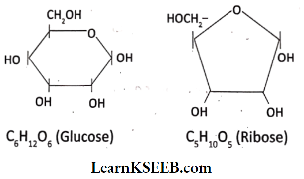
- Sugars – Example: Glucose
- Nucleotide- Example: Adenylic acid
- Fatty acids – Example: Palmitic acid
Important questions on Biomolecules Class 11 Karnataka Board
Question 4. Explain the Zwitterionic form of an amino acid.
Answer:
1. 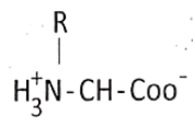 is a zwitterionic form a neutral form due to equal positive and negative charges.
2. Amino acid contains both acidic (carboxylic acid) and basic (amino group) centers and hence shows both positive and negative charges.
Question 5. What constituents of DIMA are linked by glycosidic bonds?
Answer:
- Nitrogen base and pentose (deoxy ribose) sugar, are linked by a glycosidic bond.
- This bond is formed by dehydration.
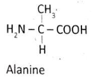
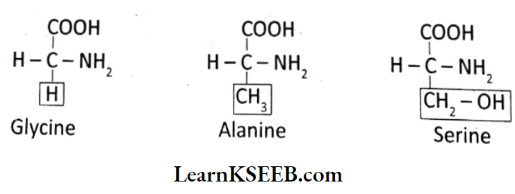
Question 6. Glycine and Alanino nro dlfforont with ronpoot to ono aubatituont on the α – carbon. What are the other common substituent groups?
Answer:
H and – CH3 are substituent groups respectively in Glycine and Alanine- at α -carbon.
Both contain- H, – COOH, and- NH2 substituent groups common
Question 7. In Starch, Cellulose, Glycogen, and Chitin are polysaccharides found among the following. Choose the one appropriate and write against each.
Answer:
- Cotton fibre _____________
- Exoskeleton of cockroach _____________
- Liver _____________
- Peeled potato _____________
Answer:
- Cotton fibre- Cellulose
- Liver- Glycogen
- Exoskeleton of cockroach- Chitin
- Peeled potato- Starch
Question 8. Find out and make a list of proteins used as therapeutic agents. Find other applications of proteins (For example: Cosmetics etc.)
Answer:
Proteins used as therapeutic agents are thrombin, fibrinogen, enkephalins, antigens, antibodies, streptokinase, protein tyrosine kinase, diastase, renin, insulin, oxytocin, vasopressin, etc.
Other applications: Proteins are also used in cosmetics, dairy industries, textile industries, research techniques, etc
Question 9. Draw the structure of the amino acid, alanine
Answer:
Question 10. Attempt titrating an amino acid against a weak base and discover the number of dissociating (ionizable) functional groups in the amino acid.
Answer:
The existence of different ionic forms of amino acids can be easily understood by the titration curves. The number of dissociating functional groups is one in the case of neutral and basic amino acids and two in the case of acidic amino acids.
Question 11. What are gums made of? Is Fevicol different ?
Answer:
Gums are secondary metabolites. It is made up of compounds present in plants, fungi, and microbial cells. Yes, Fevicol is different from gum. It is a synthetic resin made by polymerization manufactured by the esterification of organic compounds.
Question 12. Find out how much cellulose is made by all the plants in the biosphere and compare it with how much paper is manufactured by man and hence what is the consumption of plant material by man annually. What a loss of vegetation!
Answer:
About 100 billion tonnes of cellulose is prepared per year by the plants of the world. The increase in industrialization increased the use of paper. Due to this vegetation is being lost to a great extent.
Question 13. All life forms exhibit “Unity in diversity” – Give reasons.
Answer:
There is a wide diversity of all living organisms but their chemical composition and metabolic reactions appear to be similar. The most abundant chemical in all life forms is water. Living organisms contain more carbon, hydrogen, and oxygen than animate matter
Class 11 Biology Biomolecules question bank
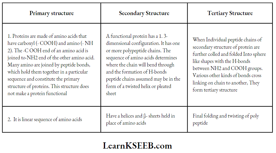
Question 14. What in meant by the tertiary structure of proteins?
Answer:
When a long protein chain of secondary structure is folded upon itself like a hollow woolen ball, It gives rise to a tertiary structure. Tertiary structure is necessary for many biological activities of protein. This gives us a 3 – dimension view of a protein.
Karnataka Board solutions for Class 11 Biology Chapter 9 Biomolecules Short Question And Answers
Question 1. Explain briefly the metabolic basis for ‘living’.
Answer:
1. Metabolic pathways can lead to a more complex structure from a simpler structure.
For example:
- The formation of sucrose from water and CO2 in the mesophyll. They are called biosynthetic or anabolic pathways.
- Some metabolic pathways may lead to a simpler structure from a complex structure.
- For example: Glucose becomes lactic acid in our skeletal muscle. They. are called degradative or catabolic pathways.
- Anabolic pathways consume energy. For instance, the assembly of a protein from amino acids requires energy input.
- Catabolic pathways lead to the release of energy. For instance, the glycolytic pathway leads to the formation of lactic acid from glucose and releases energy.
- It consists of 10 metabolic steps.
- Living organisms have learned to trap the energy liberated during degradation and store it in the form of chemical bonds.
- This stored bond is utilized as and when biosynthetic, osmotic, and mechanical work is performed.
- The most important form of energy currency in living systems is the bond energy in a chemical called Adenosine Triphosphate (ATP).
Question 2. Is rubber a primary metabolite or a secondary metabolite? Write four sentences about rubber.
Answer:
- Rubber is a secondary metabolite.
- Metabolic products that do not have identifiable functions in the host organisms are called secondary metabolites.
- Rubber is produced from the latex of Hevea and Ficus elastica.
- Latex is produced in a special type of tissue called laticiferous tissue.
- The tires of vehicles are made from vulcanization rubber.
Question 3. Schematically represent primary, secondary, and tertiary structures of a hypothetical polymer using protein as an example.
Answer:
Primary, Secondary, and Tertiary structures:
Biomolecules Questions For Class 11
Question 4. Comment on the statement “living state is a non-equilibrium steady state to be able to perform work.”
Answer:
- A living organism consists of tens and thousands of chemical compounds called metabolites or biomolecules.
- Biomolecules are present at concentrations characteristic of each of them.
- For example: The blood concentration of glucose in a normal healthy individual to 4.5 to 5.0 mm, while that of hormones would be nanograms / m/.
- All living organisms exist in a steady state characterized by concentrations of each of these biomolecules, that are in a metabolic flux.
- Any chemical or physical process moves spontaneously to equilibrium. The steady state is a non-equilibrium state. As per physics, the systems at equilibrium cannot perform work.
- Living organisms work continuously, they can not afford to reach equilibrium. Hence the living state is a non-equilibrium steady state to be able to perform work.
- The living process is a constant effort to present falling into equilibrium, which is achieved by energy input.
- Metabolism provides a mechanism for the production of energy. Hence the living state and metabolism are synonymous. Thus, without metabolism there can not be a living state.
Question 5. The dynamic state of body constituents is a more realistic concept than the fixed concentrations of body constituents at any point in time Elaborate.
Answer:
Living organisms like simple bacterial cells, protozoans, plants or animals contain thousands of organic compounds, the biomolecules.
- The biomolecules are present in certain concentrations, and ore is expressed as mols /cell or mols /liter, etc.,
- All the biomolecules have a turnover. It means that they are constantly being changed into some other biomolecules and are also made from some other biomolecules.
- This breaking and making is through chemical reactions that are called metabolism.
- Each of the metabolic reactions results in the transformation of biomolecules.
For example – The removal of CO2 from amino acids makes an amino acid into an amine.
- The majority of these metabolic reactions do not occur in isolation but are always linked to some other reactions.
- It means metabolites are converted into each other in a series of linked reactions called metabolic pathways.
- The metabolic pathways are either linear or circular, they may criss-cross each other. The flow of metabolites through the pathway has a definite rate and direction.
- The metabolite flow is called the dynamic state of body constituents. Interlinked metabolic traffic is very smooth and without a single reported mishap for healthy conditions.
- Every chemical reaction in a metabolic pathway is a catalyzed reaction. There is no uncatalyzed metabolic conversion in living systems.
- Proteins with catalytic power are named enzymes. They hasten the rate of a given metabolic conversion.
Question 6. What are macromolecules? Give examples.
Answer:
- Macro molecules are large-sized biomolecules that have high molecular weight, lower solubility, and complex molecular structure.
- It occurs in the colloidal state.
- Macromolecules are formed by the polymerization of a large number of macromolecules.
- They belong to four classes of organic compounds- carbohydrates, lipids, proteins, and nucleic acids.
Question 7. Proteins have primary structure. If you are given a method to know which amino acid is at either of the two termini (ends) of a protein, can you connect this information to purify or homogeneity of a protein?
Answer:
- The primary structure of protein is based on the number type and order of amino acid present in the chain.
- A protein has a linear structure in which the left end if the line represents the first and the right end represents the last amino acid.
- The number of amino acids in between the two ends determines the purity or homogeneity of proteins.
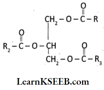
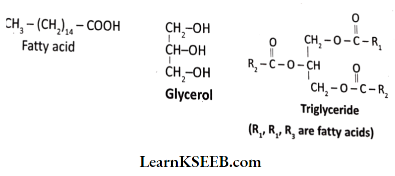
Question 8. Explain the composition of triglyceride.
Answer:
- The components of triglyceride are a single molecule of glycerol and 3 fatty acids. In glycerol 3 carbon atoms are present along with 30 n groups.
- Fatty acids consist of long-chain hydrocarbons with a carboxylic group at one end.
- Both of them form an ester bond. This bond is saturated when single-bonded carbons are present and unsaturated when double-bonded carbon atoms are present.
Question 9. Can you describe what happens when milk is converted into curd or yogurt, based on your understanding of proteins?
Answer:
- Milk Is converted Into curd or yogurt due to denaturation of proteins.
- The configuration of protein is lost. In denaturation disruption of bonds that maintain secondary and tertiary structure leads to the conversion of globular proteins into fibrous proteins.
- This involves a change in the physical, chemical, and biological properties of protein molecules.
Karnataka Board Class 11 Biology Chapter 9
Question 10. Can you attempt building models of biomolecules using commercially available atomic models (Ball and Stick models)?
Answer:
Yes, models of biomolecules can be prepared using commercially available atomic models.
- Ball and stick models and space-filling models are 3D models that serve to display the structure of chemical products and substances or biomolecules.
- With ball and stick models, the centers of the atoms are connected by straight lines which represent covalent bonds. Double and triple bonds are often represented by springs.
- The bond angles and bond lengths reflect the actual relationship. The space occupied by the atoms is either not represented at all or only denoted essentially by the relative sizes of the sphere.
Long Answers Questions On Biomolecules For Class 11
Question 1. What are secondary metabolites? Enlist them indicating their usefulness to man.
Answer:
Metabolic products that do not have identifiable functions in the host organism are called secondary metabolites. Secondary metabolites are alkaloids, flavonoids, rubber, essential oils, antibiotics, colored pigments, scents, gums, spices, etc. Many of these secondary metabolites are useful to man.
- Pigments – Example: Carotenoids, Anthocyanins etc.
- Alkaloids – Example: Morphine, Codeine etc.
- Terpenoids – Example: Monoterpenes, Diterpenes etc.
- Essential oils – Example: Lemon, grass oil etc.
- Toxins – Example: Abrin, Rlcin
- Drugs – Example: Vlnblastln, Curcumin, etc.
- Polymeric substances – Example: Rubber, gums, cellulose, etc.
Question 2. What tiro the processes used to analyze the elemental composition, organic constituents, and inorganic constituents of living tissue? What are the inferences on the most abundant constituents of living tissue? Support the inferences with appropriate data.
Answer:
To analyze elemental composition, organic constituents, and inorganic constituents of living tissue one has to perform chemical analysis.
Analyse Organic Compounds:
- Take any living tissue (a vegetable or a piece of liver etc) and grind it in trichloro acetic acid (Cif. COOH) using a motor and a pestle.
- We obtain a thick slurry.
- If we were to strain this slurry through a cheesecloth or cotton, we would obtain two fractions.
- One is called the filtrate or acid soluble pool and the other is retentate or acid insoluble fraction.
- To identify a particular compound, one has to use various separation techniques and separate an organic compound from the rest.
- Analytical techniques when applied to the compound give us the molecular formula and probable structure of the compound.
- All the carbon compounds that we get from living tissues can be called biomolecules.
Analyse inorganic compounds:
- One weighs a small amount of a living tissue (say a leaf or liver and this is called wet weight) and dry it.
- All the water evaporates.
- The remaining material gives dry weight.
- Now if the tissue is fully burnt, all the carbon compounds are oxidised to gaseous form and are removed.
- The remaining is called ash.
- This ash contains inorganic elements like calcium, magnesium, etc.
- Inorganic elements are also in acid solution fraction.
Elemental analysis:
- The elemental analysis gives the elemental composition of living tissues in the form of hydrogen, oxygen chlorine, carbon, etc.
- Analysis of compounds gives an idea of the kind of organic and inorganic compounds.
Question 3. Nucleic acids exhibit a secondary structure. Describe through Watson Crick Model.
Answer:
- Nucleic acid exhibits a wide variety of secondary structures.
- For example, one of the secondary structures exhibited by DNA is the famous Watson-Crick Model.
- According to this model, DNA exists as a double helix. The two strands of polynucleotides are anti-parallel i.e., run in the opposite direction.
- The backbone is formed by the sugar-phosphate sugar chain.
- The nitrogen bases are projected more or less perpendicular to the backbone but face inside.
- Adenine (A) and Guanine (G) of one strand pair with Thymine (T) and Cytosine (C) respectively, on the other strand. Each step is represented by a pair.
- Coiling occurs at an angle of 360°. At each step, the turn is 36°. One full turn of the helical strand involves 10 base pairs.
- The length of each turn is 34 Å
- The distance between the two steps is 3.4Å
- This form of DNA with the above features is called B- DNA
Watson and Crick’s double helix model of DNA:
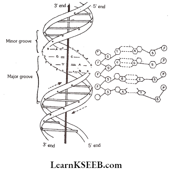
P = Phosphate, S = Sugar, A = Adenine, T = Thymine, G = Guanine, C = Cytosine
Karnataka Board Biology important questions on Biomolecules
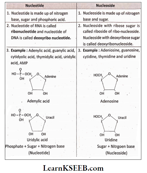
Question 4. What Is the difference between nucleotide and nucleoside? Give two examples of each with their structure.
Answer:
Question 5. Describe various forms of lipids using a few examples.
Answer:
- Lipids are water-insoluble.
- Lipids could be simple fatty acids.
- A fatty acid has a carboxyl group attached to an R-group.
- The R – group could be methyl (-CH3) or ethyl (- C2H5) or a higher number of CH2 groups (1 carbon to 19 carbons).
For example – Palmitic acid has 16 carbons including carboxyl carbon.
- Arachidonic acid has 20 carbons including carboxyl carbon.
- Fatty acids could be saturated (without double bond) or unsaturated (with one or more C = C double bonds)
Simple lipid Is glycerol which Is trihydroxy propane.
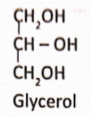
Many lipids have both glycerol and fatty acids. Here the fatty acids are found esterified with glycerol. They are then called monoglycerides, diglycerides, and triglycerides.
Triglyceride (R1, R2, R3 are fatty acids):
- These are also called fats and oils based on melting point.
- Oils have lower melting points
Example: Gingely oil and hence remains as oil in winter.
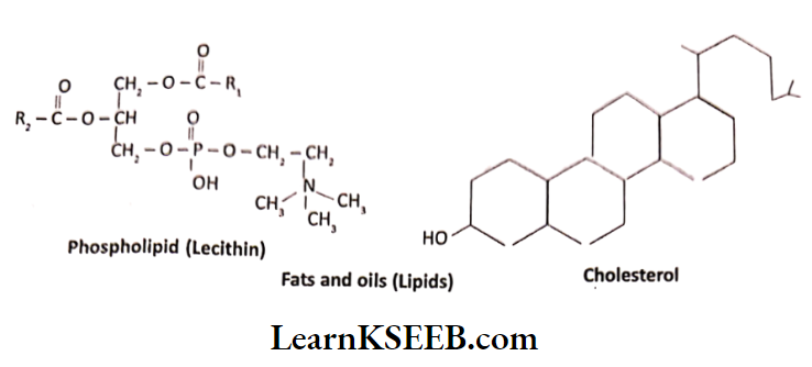
- Some lipids have phosphorous and a phosphorylated organic compound in them. These are called phospholipids. They are found in cell membranes.
- One example is Lecithin.
- Some tissues especially the neural tissues have lipids with more complex structures.
- If a phosphate group is also found esterified to the sugar, they are called nucleotides.
Examples: Nucleotides are adenylic acid, thymidylic acid, guanylic acid, uridylic acid arid cytidylic acid.
Question 6. Illustrate a glycosidic, peptide end n phoepho-diostor bond.
Answer:
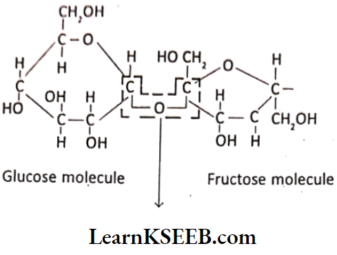
Glycosidic bond:
In a polysaccharide, the Individual monosaccharides are linked using a glycosidic bond. This bond Is formed by dehydration. This bond is formed between two carbon atoms of two adjacent monosaccharides.
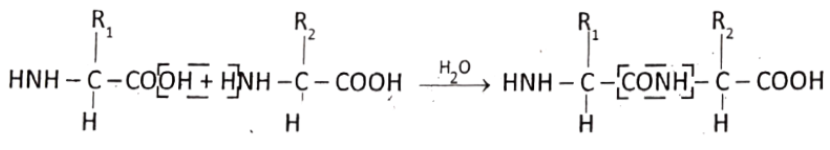
Peptide bond:
In a polypeptide or a protein amino acids, are linked by peptide bonds. These bonds are formed by the reaction between the carboxyl group (- COOH) of one amino acid with the amino group (NH2) of the next amino acid, with the elimination of water.
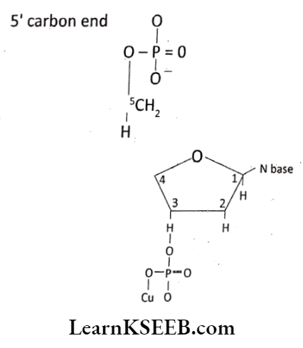
Phospho-diester bond:
In a nucleic acid a phosphate moiety links the 3′ – carbon of one sugar of one nucleotide to the 5′ carbon of sugar of the succeeding nucleotide. The bond between the phosphate and hydroxyl group of sugar is an ester bond. As there is one such ester bond on either side, it is called a phospho-diester bond.
Question 7. Find and write down structures of 10 interesting small molecular weight biomolecules. Find if there is any industry that manufactures the compounds by isolation. Find out who are the buyers.
Answer:
1. Sugar (Carbohydrates):
Amino Acids:
Fats:
Oils:
Question 8. Find out qualitative test proteins, fats, oils, and amino acids test any fruit juice, saliva, sweets, and urine for them.
Answer:
Biuret test for protein:
The biuret test Is a chemical test used for determining the presence of peptide bonds, In a positive test, a copper II ion (Cu;‘ Ion) Is reduced to copper (Cu’) which forms a complex with the nitrogen and carbon of peptide bonds in an alkaline solution. A violet color indicates the presence of protein.
Ninhydrin test for amino acid:
- Ninhydrin is a chemical used to detect ammonia or primary and secondary amines.
- When reacting with these free amines, a deep blue or purple color known as Ruhemann’s purple is evolved.
- Most of the amino acids are hydrolyzed and reacted with ninhydrin except proline (a secondary amine).
Solubility test for fats and oils:
A positive solubility test for fats is that the fat dissolves in lighter fluid and not in water. In this test, 5 drops of fat or oil are added in two test tubes containing 10 drops of lighter fluid and 10 drops of cold water respectively.
- Fruit juice: Fruit juice contains sugar. So it cannot be tested by the above-mentioned test.
- Saliva: Saliva contains proteins, mineral salts, amylase, etc. So it can be tested for proteins and amino acids.
- Sweat: Sweat contains NaCl salts.
- Urine: Urine contains proteins. So it can be tested for proteins.